Live from Prospect St: The Big Crunch (part 2)

Continued from Part 1

Since the two children appear generally intact, you ask your partner to evaluate them more fully while you head for the sedan to find the driver. Anticipating three transports, two stable and one potentially critical, you ask your dispatch to continue the P12, and also to ensure that police are en route (they are).

Arriving at the sedan, you find a middle-aged woman in the driver’s seat, alert. She is pink and warm, perhaps more diaphoretic than you’d expect for the ambient temperature, and does not initially notice as you kneel beside her. A firefighter is holding C-spine immobilization from the back seat.

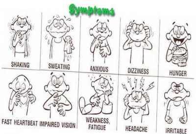
When you greet her and pat her on the shoulder, she gives no response, but with more vigorous stimulation she looks over and acknowledges you distractedly. With multiple attempts and some yelling, you’re able to get answers to a few questions, but she is slow, tangential, and often ignores you outright. She gives her name as Samantha, but cannot or will not provide her last name; she is unable to describe the events that led to the collision; and she gives no medical history or current medications. She does state several times that she’s fine and would like to leave. When asked about her passengers, she mumbles “my kids” and mentions her brother several times. She endorses pain when asked explicitly, but does not specify where. She agrees that she drank “a little” alcohol; when asked about any drug use, she denies it vehemently.

Physically, she appears generally unremarkable. She is breathing somewhat shallowly but effectively, and her radial pulse is around 100 and slightly weak. Her seatbelt is not in place, but it’s unclear whether it was removed at some point. No gross trauma is apparent upon her head, face, or neck, and she does not complain or grimace upon palpation. She is uncooperative with a neurological exam, but demonstrates spontaneous movement of all four extremities. Her pupils are equal and seem appropriately small on this moderately bright day. Chest rise is generally equal and her abdomen is supple; no bruising consistent with seatbelt injury is visible. Her left knee is abraded and somewhat swollen. A sprinkling of dark blotches and streaks are noted on her left ventral arm in the antecubital region. Both frontal airbags are deployed; the windshield is cracked, but lacks a “starred” point of impact; and the plastic dashboard in the driver’s knee area is damaged and cracked. No blood or other damage is visible in the interior compartment. There are no child seats.

Your partner comes over. “The kids seem fine, just upset. One’s complaining of some abdominal pain, but it looks okay. They’re little troopers. Fire says they were wearing regular lap belts with the shoulder strap tucked behind them.”

When you wonder aloud whether there are more patients, he says, “There was nobody else in the car when fire arrived. The truck driver gave a statement to the police about how she was swerving across the road and plowed into him, but then he eloped.” He looks over your shoulder. “Oh, and the P12 is pulling up now.”


What is your treatment plan for these three patients? What are their respective priorities, any points of concern, and how could you shed additional light on their status?

Who will transport which patient, and to which destinations?

What special considerations should be made during documentation?


The conclusion is here

Live from Prospect St: The Big Crunch (part 1)

It’s 4:00 PM on a gloomy Friday in Chandlerville, and you’re the technician for the A2, a dual-EMT, transporting BLS unit dedicated to the city. Chandlerville is a small town, but densely populated, and its numerous industrial districts are frequent sources of work. 911 dispatch is directly through the fire department, which also sends a BLS fire apparatus to assist on all medical calls; your company’s ALS is also available by request. You are equipped with finger-stick glucometry, glucose, aspirin, and epinephrine.

After a “man down” call that ended in a patient refusal, you’re now returning to quarters. Just as you’re beginning to back into the garage, a tone sounds.

Engine 3 and Ambulance 2, respond to 2108 Coastal Rd, the Empire Shipping Company, for an MVA. That’s two-one-oh-eight Coastal Road, in front of Empire Shipping, for an MVA. Engine 3?

“Engine 3 is responding.”

Ambulance 2?

As your partner flips on the lights and pulls out to the street, he speaks into the radio: “Ambulance 2 has 2108 Coastal Rd.”

Time out 16:01.

Coastal Road is a long connector that wraps around the edge of town, and you glance at the map book to confirm that the 2000 block will be near the very end, about as far away as you can get in Chandlerville. Engine 3 is stationed in that district, however, so they arrive within minutes.

“Engine 3 to Firecom.”

Firecom answering.

“We’re off at 2108 Coastal Road. Two-car MVA, car versus truck. Multiple injured parties and entrapment. Start an ALS unit and a ladder for extrication.”

Engine 3, you have a car versus truck, multiple injuries with entrapment. Break. Ladder 3, respond to 2108 Coastal Rd for the MVA; Engine 3 is on scene and A2 is responding. Time out 16:04.

A few seconds later, your company radio dispatches Paramedic 12 to the same address, after Chandlerville Firecom contacts them via landline. The P12 starts responding, but they’re coming from two towns away, with an ETA of 10+ minutes. The field supervisor also starts rolling from an unknown location to assist. 30 seconds later, Engine 3 updates that they have an injured adult and several children.

Now very awake, you reflect that the nearest hospital will be Chandlerville Memorial, a 3–5 minute emergent transport (10 minutes otherwise). The nearest large tertiary center, Bullitt Medical Center — a Level I adult trauma center and a designated pediatric ED — is 15 minutes emergently (25 otherwise). The nearest Level I pediatric trauma center, however, is the Children’s Hospital, which is also 15 minutes but in the opposite direction; they do not receive adult patients.

Ladder 3 arrives on scene momentarily, and you pull up a few minutes later. As you park and call yourself out, you observe a Ford sedan with its front left corner smashed in, two feet of its fender and frame crumpled. This is evidently the result of driving almost headlong into the side of an 18-wheeler. It appears that the driver swerved right to avoid the truck, undercutting its rear wheels and “submarining” itself; the damage reaches the passenger compartment, but there does not appear to be significant intrusion. The truck itself seems minimally damaged.

As you jump out, a firefighter waves you down. “We’ve got three!” he announces. “Mom’s in the driver’s seat; she seems really loopy, probably drunk. Her door is just dented, we popped it open. But her kids are over there.”

Twenty feet away, you see two young girls, around 4 years old, each in the arms of a firefighter. They are crying loudly and clearly upset, with no visible injuries. The mother is hidden from sight in the sedan. The driver of the truck is nowhere to be seen.


What are your initial steps for addressing this scene?

Who appears to be the first priority for care?

What resources will you need? Which, if any, should you cancel?


Continued in part 2 and the conclusion

The Curious Incident of the Dog in the Diagnosis


“Is there any point to which you would wish to draw my attention?”

“To the curious incident of the dog in the night-time.”

“The dog did nothing in the night-time.”

“That was the curious incident,” remarked Sherlock Holmes.

Sir Arthur Conan Doyle, Silver Blaze

We can learn a lot from Sherlock Holmes.

If there’s anybody who better personifies the ultimate diagnostician, I don’t know who. Sir Arthur Conan Doyle, creator of the Holmes canon, was himself a physician and purportedly based his famous detective on Dr. Joseph Bell — who, it was said, could glean a dozen esoteric facts of a patient’s background, history, and complaints from a single glance. (Holmes himself, of course, was not a medical man; that role was played by Watson, the earnest physician who carried his stethoscope wrapped inside his hat.)

Holmes didn’t diagnose illness. Instead, he diagnosed crimes. But the methods were the same, so much so that among the countless fictional characters based upon the Holmesian archetype, some have been crime-solvers (cf. Monk), yet others have been medical doctors (House is the best). Perhaps we shouldn’t model ourselves after the man, who was a single-minded addict and misanthrope, but when it comes to diagnosis — something we can’t escape in medicine — he knows whereof he speaks.


The diagnostic method

Holmes tells us in The Sign of Four that detection involves nothing more than three skills: observation, deduction, and knowledge. Let us consider what he means when faced with, for example, a complaint of chest pain.

Observation: we perceive a middle-aged male, alert and seated upright, rubbing at his sternum with a pained expression. His skin is slightly pale, his respiratory rate is slightly elevated, and he is hypertensive. He complains of “tight” 4/10 chest pain whenever he breathes. Upon auscultation we detect diffuse, bilateral, biphasic wheezing. We note a history of coronary artery disease, diabetes, and COPD.

Knowledge: Chest pain in adults indicates a high risk for acute coronary syndromes. Pallor, tachypnea, and hypertension are consistent with this diagnosis. Sharp, mild, pleuritic pain is not, nor is wheezing, all of which are more consistent with a primarily respiratory etiology. But we also know that MI often presents atypically, particularly in diabetics.

Deduction: Both cardiac (ACS) and respiratory (COPD exacerbation) diagnoses top our differential. An ECG and biomarkers are needed to further evolve the odds.


So what just happened? We observed using our medical assessment — the history, physical, and diagnostic tests — thus yielding a collection of facts and data. We took the set of background knowledge we already possessed, regarding pathophysiology, epidemiology, and hazard ratios, and used it to “fill in the blanks” and provide context to our assessment findings. Finally, we connected the dots together and used deduction to decide what we’re dealing with.

Holmes knew this method well. He might observe your tattered boots, and using knowledge he possessed of typical wear patterns in the various trades, deduce that you make your living as a longshoreman. Simplicity itself.

Why is this a useful model for diagnosis? Because it highlights the fact that these three skills are entirely distinct, though all quite essential. Observation requires skill with the physical exam, the ability to take a nuanced history, the acumen to interpret diagnostics — it’s simply the trait of being aware. (Holmes, succinctly: “Data, data, data! I can’t make bricks without clay.”) Knowledge is knowledge: it’s memorized facts, what you learn in school or from books, and it gives us the basis to understand the raw material we discover in our assessment. Finally, deduction is the mental capacity to analyze, discover patterns, weigh odds, use your imagination, and extract from the vast pool of observation and knowledge the particular pieces that are actually relevant. (Holmes: “… to recognize out of a number of facts which are incidental and which are vital.”)


The hidden danger

Here’s the rub: we’re almost too good at deduction. Humans are excellent at finding patterns in anything. If I leave you my tea-cup for long enough, you’ll undoubtedly find an image revealed in the leaves.

That’s good — but it’s an error. Because there’s not really any image in the tea leaves. But if you’re good at observing details, and have a strong imagination, you’ll still “deduce” many wonderful things from it. Call it apophenia: people want to connect the dots, even when there aren’t any. So we create connections that may be true, but are not always true. We develop stereotypes. Simplifications. False associations.

In medicine, we’re especially prone to this. Because we do know that the human body is interconnected, and that patterns are the rule rather than the exception. Indeed, a large part of developing experience and clinical judgment is increasing your catalog of mental connections. Crackles mean CHF. Irregular pulses mean A-fib. People with Foley catheters have UTIs. Homeless people are drunk. Toe pain is a nonsense complaint. We can’t avoid making the connections, because just like when Holmes examines your boots, those connections are essential to doing our job. But at the same time, we need to learn when to reign them in, or we enter an inescapable diagnostic tunnel after the first moments of patient contact.

It is a capital mistake to theorize before one has data. Insensibly one begins to twist facts to suit theories, instead of theories to suit facts.

A Scandal in Bohemia

What’s the secret? Knowledge.

Sure, we gather the pieces from our assessment, and we automatically start to connect them together. We can’t help that; patterns jump out at us, we’re natural pattern-finding machines. But using our knowledge, we can look past those simplistic, eye-catching patterns, because knowledge tells us something more subtle: what’s missing.

She’s all false positives. See, that’s the trouble with naturals. They don’t see what’s missing.

Lie to Me, “Moral Waiver”

Okay, Friday night, a “man down” call for a homeless guy on the sidewalk. You’re already thinking: drunk. And the initial observations confirm it: he rouses sluggishly, slurs his speech, and pushes you away as he rolls back over. But then you open the mental box that you filled with this sort of thing in your training, and you reflect: where’s the bottle? And this is a strange spot — it’s cold, wet, public and unsheltered. And come to think of it, is that a medical alert bracelet? We should probably check this guy out a little more. Maybe take his blood sugar, look for any trauma, shake him awake and ask some questions.

The initial pattern recognition is there, but you don’t have to be a slave to it, because you know what else to look for. Even if five clues say one thing, if we don’t see five others that ought to be there, that tells us something different. Pertinent negatives, they call ’em in the business.

Maybe there’s nothing else; maybe the drunk is just drunk. But we’re too smart to make that kind of assumption. Because we know that getting it right doesn’t just mean registering the hits — it means checking off the misses, too.

Holmes would expect no less.


Glucometry: Clinical Interpretation

Continued from Glucometry: Introduction and Glucometry: How to Do it

Implementing glucometry into your overall assessment means understanding three things: when to use it, what the results mean, and when it fails.


Indications

First of all, by and large the only people with derangements of their blood sugar should be diabetics. The rest of us are generally able to maintain euglycemia through our homeostatic mechanisms, except perhaps in critical illness causing organ failure and similar abnormal states. Now, if someone injected you — a non-diabetic — with a syringe of insulin, you’d become terribly hypoglycemic, since it would overwhelm your body’s ability to compensate for the loss of glucose. But nobody’s likely to do that if you’re not a diabetic, unless it’s meant for somebody else and a drug error occurs, or I suppose if they’re trying to assassinate you.

With that said, people walk around who are diabetic and don’t know it. I’ve lost track of the patients I’ve transported who presented with signs suggestive of a diabetic emergency, denied a history of diabetes, and came back with a BGL of 600. Well, my friend, I have some bad news for you. “Everybody is diabetic, even if they’re not” is my attitude. Almost a fifth of older Americans are diagnosed, and the older and sicker they are, the more common it is.

Which brings us back to: who needs a BGL?

The most correct answer is anybody with clinical indications of either hypo- or hyperglycemia. As we saw, diabetes itself is really associated with hyperglycemia, which is why the classic signs of hyperglycemia are usually used to diagnose diabetes: polyuria (excessive urination, as extra glucose is excreted by the kidneys and brings water along with it osmotically), polydipsia (excessive thirst and water consumption, to replace the fluids urinated out), and polyphagia (constant hunger, since despite all the sugar floating around it’s not reaching the cells very easily). If your patient is complaining of those, you might be the first one to discover their condition. The diagnosis doesn’t require elaborate tests and imaging; a fasting glucose over 126 BGL tested on multiple occasions, or just once in combination with clinical symptoms, or a post-prandial (after eating) glucose exceeding 200, is the definition of type II DM. (With that said, I wouldn’t go around diagnosing your patients; that’s not your job, and you’re not quite that good.)

Once the glucose gets higher than the “renal threshold” — usually around 180 in average folks — the body starts to excrete it into the urine. This can actually be detectable by chemical dip-stick, or even by odor and texture at very high levels.

When hyperglycemia becomes severe and prolonged enough, we start to worry about diabetic ketoacidosis. Although burning fat and protein is not necessarily dangerous (some popular diets actually put you into a mild ketogenic state intentionally), extensive accumulation of ketones caused by a total lack of insulin (as in type I diabetics — DKA is rarely seen in type II) creates a metabolic acidosis in the body. This is when the long-term harm of hyperglycemia becomes a short-term hazard. DKA causes altered mental status, usually elevated states of confusion and disorientation, and combative behavior isn’t uncommon. Combined with the acetone odor that sometimes presents on the patient’s breath — which can smell like alcohol — DKA patients can seem suspiciously like drunks, and treating them like drunks is a great way to go down a bad path. (A word of wisdom: not only is everybody diabetic, but drunks are definitely diabetic.) DKA also frequently presents with symptoms of dehydration, due to the osmotic water loss in the urine; nausea and vomiting; and deep, rapid Kussmaul breathing to blow off the acidic CO2.

A few situations can cause short-term hyperglycemia, including stressors of any kind (there’s even “white coat hyperglycemia,” where patients tend to produce elevated sugars at the doctor’s office), but these typically won’t produce anything like the massive levels leading to DKA.

With all of that said, you need to really build up some glucose before hyperglycemia becomes symptomatic, and even more than that before it becomes acutely dangerous and unstable. That’s why as a rule, we’re more concerned with hypoglycemia, usually due to medication administration, physical exertion, or metabolic demand exceeding what was expected. Hypoglycemia again presents as altered mental status, in this case more often an inhibited rather than an elevated state: confusion, lethargy, disorientation, inability to focus or follow commands, weakness, headache, seizures, and eventually coma and death. The fun part is that the impairments can present as focal as well as generalized deficits: unilateral weakness of the limbs or face, speech slurring, poor gait, vision abnormalities, and more. In fact, hypoglycemia is a neurological chameleon, and can look like almost anything; it’s particularly notorious for imitating strokes, and for causing (not imitating) seizures. Interestingly, kids are particularly prone to hypoglycemia due to their gigantic heads, full of glucose-hungry brain.

Despite all this, the primary manifestations of early hypoglycemia are actually not symptoms of hypoglycemia. Rather, they’re caused by catecholamines — by the body releasing stress hormones, primarily epinephrine, in a response to the emergency. (This is not an irrational move: epinephrine helps us release and retain glucose.) As a result, we often seen the same signs we’d expect in anybody with a profound sympathetic stimulus: pale and diaphoretic skin, anxiety and shakiness, tachycardia and hypertension, even dilated pupils. Wise diabetics recognize the early signs of this sympathetic response and drink some Pepsi. As levels keep dropping, these symptoms combine with the neurological effects of glucose starvation to produce a confused, sweaty, increasingly stuporous individual. If left untreated, finally the sugar drops until we’re looking at the picture of impaired and diminished consciousness caused by true hypoglycemia. So just like always, the signs of compensation are our early warning system; once the body decompensates, it’s already late in the game.

To make a long story short, anybody with altered mental status, or any kind of general systemic complaint (weakness, fatigue, anxiety, nausea, etc.) should probably get their glucose tested, whether or not they have a known history of diabetes. This is true even if you suspect another cause, such as stroke. Not only can diabetic emergencies look like anything, they can also be comorbid; it is extremely common for patients to have another problem, yet also to bring a high or low sugar along for the ride, due to the illness throwing a wrench in their normal intrinsic and extrinsic glycemic homeostatic systems.

A number of years ago, there was some limited but compelling research that suggested poorly-controlled blood glucose (meaning not severe derangements but merely small deviations from the ideal range) was associated with increased mortality among an inpatient population with a wide variety of conditions. In other words, if you were hospitalized with something like sepsis, you were more likely to end up dying if your sugar tended to float around 160 instead of 110. As a result, it become trendy to practice extremely tight and aggressive glucose management for virtually everybody; diabetic patients were being tested every few hours and ping-ponged around using medication to keep their numbers textbook-perfect. More recently a number of studies have suggested that this may be less important than was thought, and in fact that excessive paranoia leads to a lot of iatrogenic harm from accidental insulin overdoses. This battle is still being fought in the hospitals, but for our purposes a reasonable take-away would be: when managing acute illness, from sepsis to head injury to cardiac arrest, once everything else is done it’s not a bad idea to check the patient’s sugar.


What’s the Number Mean?

So you’ve taken a blood glucose, either by capillary finger-stick or from a venous sample. Now what?

We mentioned that the “normal” range is something like 70–140. Diabetics seeking to control their condition and not have their toes falling off in a few years usually strive for tighter control of their BGL than is needed for acute care; a sugar of 175 is a little on the high side for a routine check, but a pretty meaningless elevation for our purposes.

All things are also relative, in that a given BGL must be compared to the patient’s baseline to predict its effects. In other words, poorly-controlled diabetics who are routinely sitting at 200 may become symptomatic of hypoglycemia at relatively high levels, whereas very well-controlled diabetics who usually run lower may be able to drop very low indeed without noticing it. However, a few rules-of-thumb are useful:

Non-diabetics usually become noticeably symptomatic below a sugar of, on average, about 53. (Diabetics, particularly those who are usually poorly-controlled, are more variable — their average symptomatic threshold is more like 78.)

After a recent meal, diabetics may demonstrate hyperglycemia to various degrees depending on whether they ate a Cobb salad or an entire chocolate cake. Non-diabetics should not exceed 200 or so. A few people can exhibit hypoglycemia after meals, due to alcohol consumption, “dumping syndrome,” or some other phenomena, but far more often they’ll exhibit similar symptoms without any true hypoglycemia; some people get shaky and sick due to postprandial epinephrine release.

After an unusual period of fasting (“haven’t eaten since yesterday”), non-diabetics should still have a largely unremarkable sugar. For diabetics, it will depend mainly on how much and what type of medication they’re using.

There’s usually a gap of 10–20 mg/dL between hypoglycemia that’s noticeable to the patient (i.e. sympathetic effects) and hypoglycemia that causes cognitive impairment (i.e. neurological changes). This is their safety margin, when they’re taught to eat or drink some fast carbs; if it keeps dropping they may no longer be able to take care of themselves.

But here’s the problem: the sympathetic “warning signs” can be mediated or impaired for various reasons. For one thing, if your body has to flip that switch often, you become numbed to it, and your hypoglycemic thresholds becomes lower and lower. And many patients with various metabolic and endocrine failures simply can’t muster much of a stress response — the same reason why the elderly may not produce tachycardia and other shock signs when they become hypovolemic. Finally, drugs like beta blockers that directly block sympathetic activity can seriously obscure hypoglycemia. Grab your nearest bottle of beta blockers and read the list of adverse effects: one will be hypoglycemic unawareness, a five-dollar term that means beta blockade can make it difficult to know when your sugar drops low.

Another important consideration in evaluating glucose levels is the expected trend. For instance, a BGL of 70 in a diabetic patient might not excite anybody. However, if you’re testing her because her nurse said that she just accidentally received four times her normal insulin dose, then a BGL of 70 should be alarming, because it’s probably going to keep dropping, and she doesn’t have very far to go.

To make a long story short, the clinical effects of both hypo- and hyperglycemia can vary substantially. What to do? It’s simple: assess the patient physically, obtain a history of their oral intake, medications, and metabolic demands (such as exercise), test their sugar if there’s any possibility of glucose derangement, and compare all those data against each other. A low number in the setting of obvious clinical symptoms is bad. A low number in an asymptomatic patient, or a normal number in a patient with highly suggestive signs and symptoms, should force you to bring out your thinking cap and weigh the odds.

What about treatment? Severe hypoglycemia needs ALS or the hospital — they’ll receive IV dextrose. Severe hyperglycemia needs the hospital only, where they’ll receive carefully-dosed insulin; this is generally considered too dangerous to administer in the field (although patients may have their own), so paramedics are reduced to giving fluid boluses, which may help dilute high glucose concentrations (not a very elegant solution) and is probably needed by a patient in DKA anyway, but isn’t really a fix.

What about oral glucose, in the cute little tubes we carry? Typically these are gels containing 15g of glucose, taken orally (either swallowed or held in the mouth — against the cheek or under the tongue — until it’s absorbed). Do they work? Sure. But it’s not much sugar and it’s not very fast. I found one source that suggests 15g of oral glucose should raise the BGL by 50 mg/dL within 15 minutes of administration — but I’ve never found it to be nearly that effective. In my experience, a bump of about 10 mg/dL per tube is about the best you can hope for in the short-term. If you need more than that, go with the medics and the IV syrup.


Testing Errors

When is a tested capillary or venous glucose unreliable? Usually it’s your fault.

Well over 90% of BGLs that test outside the maximum error range (remember, around 15%) are due to user error. Some of the main ones:

  • Your meter requires lot coding, and you failed to do so or used strips from the wrong lot.
  • You failed to clean the skin before lancing, contaminating the sample (not to mention creating an infection risk), or you had some D50 on your glove and it got mixed in there.
  • Rather than gently wicking the sample into the strip, you “smeared” the two together with mechanical pressure, interfering with the expected reaction process.
  • You drew blood from an arm with an IV infusion of D50, TPN, or other meds distal to it. Particularly when peripheral perfusion is poor, always try to sample at a different limb from any running drips.
  • You tried to reuse a non-reusable strip (gross).

Okay, okay, so nobody’s perfect. Factors that may not be as obvious include:

  • Temperature. The test reaction is designed to function within a specific temperature range, which is broad (often around 40–104 degrees) but not limitless, so don’t use them in freezing weather, and try not to leave your equipment ungaraged without climate control when it’s very hot or cold out.
  • Altitude. Just in case you’re an Everest expedition doctor.
  • Humidity. The strips have trouble when it gets very humid.
  • Air. The reagents in the strips will actually degrade if exposed to air for sufficient periods of time, so make sure that you keep them in their tightly-sealed case, and follow their printed expiration dates.
  • Time. If you draw whole blood and leave it around (much more likely to happen in the laboratory than in the ambulance), the erythrocytes will metabolize glucose at about 5-7% per hour.

The good news is that in many of these situations, internal error-checking within the glucometer will recognize the problem, and flash an error rather than a reading. Errors messages are usually numbered and can be informative, but each manufacturer uses different codes, so read the manual if you want to know what “ER2” means. (Hint: not enough blood in the sample is by far the most common.) Many of the other problems can be caught if you regularly check the meter using a known-value test solution, which you should be doing anyway according to most drug and safety agreements. (By the way, both the test strips and those vials of solution are usually meant to expire a few months after opening — the printed date is for an unopened bottle — so if they’ve around forever it’s probably time to retire them.)

What about physiological states that can interfere with the reading? We’ve discussed a few, but briefly:

  • Hematocrit. Anemia from any cause, including cancer or blood loss, causes falsely high readings. High crit, common in neonates, causes falsely low readings.
  • PaO2. Oxygen interferes with the electrochemical redox reaction; thus high concentrations of dissolved oxygen cause falsely low readings, and low PaO2 (i.e. hypoxia) cause falsely high readings, potentially masking a true hypoglycemia.
  • pH. Primarily in meters using the glucose oxidase enzyme, alkalosis will cause falsely elevated readings, while acidosis causes falsely low readings. The acidosis of DKA can therefore cause falsely low readings, masking the profound underlying hyperglycemia, so if the clinical picture screams DKA, don’t necessarily let the glucometer tell you different.
  • Macronutrients. High levels of circulating proteins or fats can cause falsely low readings due to dilution.
  • Hypoperfusion and inadequate circulation. See our previous remarks on this, and remember that venous sources will be more accurate than capillary.

Finally, are there medications that can interfere with glucometer accuracy? There sure are. These in particularly are highly device-dependent, with the glucose oxidase-type meters most often affected. Generally, the effects are not profound, but occasionally they may be clinically relevant.

  • Ascorbic acid. Better known as Vitamin C, some people take megadoses of this stuff, thinking it’ll cure their cold or flu. Depending on the meter it can cause falsely high or low readings, usually a minimal change, but at “megadose” levels the effect can be significant.
  • Acetaminophen. Also known as Tylenol. The effect is similar to ascorbic acid, but even more modest; it should only be considered in major overdoses, and even then the difference is unlikely to break 35.
  • Dopamine. Massive doses, such as might be used for intensive inotropic support, can modestly influence glucose dehydrogenase-based meters.
  • Mannitol. High doses can elevated the measured BGL by around 35.
  • Icodextrin. This is a dialysate solution used for peritoneal dialysis (not hemodialysis — this is where they pump fluid into the abdomen, let it sit, then drain it out), mainly in patients with diabetes. It metabolizes to maltose, which can cause falsely elevated readings in certain meters. There’s at least one tragic and unfortunate case report of a patient death resulting from massive insulin overdose due to this effect, not noticed until the true BGL was obtained by laboratory analysis. If your patient undergoes peritoneal dialysis, try to find out what dialysate is used, and if that’s not possible, it may be safest to assume their sugar is lower than you’re measuring.


Conclusions

After all this you’re probably thinking glucometry is so convoluted and rife with pitfalls that you’re better off just eyeballing how sweet your patients are. But don’t let me turn you off! This remains one of the best assessment aids we have, because diabetic emergencies remain some of the most common, most treatable, and most easily confused disorders that we encounter. We can’t perform exploratory surgery, and we may never see prehospital CT scans, but this is a diagnostic test that’s so cheap and simple, with such real potential to affect your decisions, that it should be available everywhere. If you maintain your equipment, learn how to do it right, and keep a few basic confounders in mind, it’ll serve you well as one of your most reliable tools.

Glucometry: How to Do it

Read part one at Glucometry: Introduction

So we want to know how much glucose is in our blood. How can we determine this?

Most modern systems involve a handheld electronic meter, which accepts disposable test strips. The general method:

  1. Insert a strip into the meter; this usually turns it on automatically, and the screen will indicate when it’s ready for a sample.
  2. Clean the patient’s fingertip with an alcohol swab.
  3. Using an automatic lancet (a spring-loaded needle), prick their finger-tip, drawing out a droplet of blood. You may need to push or massage the skin toward the puncture site in order to “milk” blood out, particularly if there’s poor circulation.
  4. [Optional] Many services recommend wiping away the first drop of blood and drawing out a second for your sample.
  5. Once you have a sizable, “hanging” drop of blood, apply it directly to the sample site on the test strip. It will wick inside and be absorbed.
  6. The meter will usually display some kind of count-down. Once it’s finished analyzing, it will show the blood glucose concentration (BGL) in mg/dL or mmol/L.
  7. Apply a band-aid to the site, and dispose of the test strip, lancet, and other bloody bits as appropriate.

What magic happens when you apply blood to the strip? There are a few methods.

(Skip this paragraph if chemistry wasn’t your favorite class.) As a general rule, the glucose in the sample is broken down by an enzyme (often glucose oxidase, or a version of glucose dehydrogenase). This reaction is proportional to the glucose concentration, and can be visualized by the accumulation of an indicator; the more glucose that reacts, the more color develops, and this is measured by a photometric transmission sensor. Alternately, in most current sensors, a more modern and somewhat more robust electrochemical method is used; here glucose is selectively oxidized, and electrons are pulled across a mediator to an electrode, which measures the current generated — either average, peak, or total depending on the type of analysis.


Results

Across the US, blood glucose is measured in the units mg/dL (milligrams per deciliter). In much of the rest of the world, the unit is mmol/L (millimoles per liter). This means that if your paramedic buddy from the UK is telling you about a diabetic he treated, the numbers may seem peculiarly low. Since we’re mostly Yanks here, we’ll be working in mg/dL, but if you ever need to convert to mmol/L, you can simply divide it by 18 (or multiply by 18 to get from mmol/L back to mg/dL).

Much like vital signs, textbook ranges for “normal” blood glucose levels vary. A loose range for practical purposes would be around 70–140, although ideally we should be under 100 most of the time, and routinely testing over 125 is not a great indicator for your health. Numbers will be elevated after eating, but non-diabetics still shouldn’t break 200 or so.

Although we’ll talk more about clinical interpretation later, in general it’s safe to say that the lower the number, the more each point matters. The difference between 70 to 50 can be profound, while the difference between 200 and 180 may be totally undetectable.


Accuracy and Precision

Glucometers have evolved through quite a few generations by now, and they continue to improve in robustness and reliability. Most diabetics use them regularly to track their sugar and thereby guide their diet and medications.

How accurate are they? Depends on who you ask. The American Diabetes Association says that at a minimum, they should give readings within 15% of the true value, and ideally manufacturers should shoot for an error of under 5%, at all concentrations. But percentages can be a confusing way to measure it, because as we observed, a 15% difference at a sugar of 500 (a possible range of 425–575) may mean little, while a 15% difference at a sugar of 60 (a range from 59, which is low, to 69, which is about normal) can be rather important. So the FDA says this instead: 95% of the time, for values below 100, meters should be within 20 points of the true value, while for values above 100, they only need to be within 20 percent.

Whatever the case, every meter varies, but generally they can be relied upon to fall within about 15% of reality, as long as no user errors or confounding factors (we’ll talk about those) are present.


Blood Source

Traditionally, capillary blood for glucometry is taken from the fingertips. This is painful, so most modern glucometers have been evaluated to determine their accuracy when blood is drawn from alternate sites. Any location with lean, vascular muscle close to the surface (i.e. not too much fat overlying, which you may not be able to penetrate with a lancet) can be usable — the forearm is the most common site. The research has shown that this practice is generally fairly accurate for routine purposes, but the danger is that BGL from the forearm lags behind that from the fingertips. It takes longer for these readings to approach reality — about 30 minutes, in fact, before you’ll read the same from the forearm as you’d read at the fingertip, and until then the numbers may be radically wrong (for instance, a reading of 145 when it’s really 50). So glucometer manufacturers recommend that diabetics always use the fingertip when there’s any question of hypoglycemia, when they’ve recently eaten, or any time when it’s important to have the most current and accurate figure. Obviously, this is always important for EMS, so we should generally stick to fingers.

On the other hand, in many areas it’s common for paramedics to start IVs and then use a drop of blood from the catheter’s flash chamber for glucometry. Briefly, like so:


A used catheter (needle inside)


The rubber stopper behind the flash chamber


Press on the rubber until a usable drop of blood comes out the end


This method works, saves you the trouble of lancing a finger, and spares the patient some extra pain. But it’s usually considered technically incorrect, because the blood in the catheter is venous, whereas glucometers are calibrated for capillary blood. See, since venous blood has already given up glucose to the tissues whereas capillary blood is still in the process of doing so, venous BGL is lower than from capillary sources — usually about 5–10 mg/dL. (If by chance you have a source of arterial blood, then that should be higher still.) However, after eating, particularly carb-rich foods, capillary sugar may be as much as 25% higher than venous, because of the extra glucose sequestered in the muscular tissue. (Stockpiling this fuel is why marathon runners like to “carbo load” before events.)

With that said, I’m going to make a controversial recommendation: in most cases, whenever it’s available, venous blood should be used instead of capillary blood. If someone has started an IV, then you should be using that instead of a fingerstick. Why? Despite the small and usually predictable difference, in sick people, it’s actually a more accurate result.

In sick people, circulation is often impaired; this is particularly true in situations like shock, sepsis, and the mother of all shock states, cardiac arrest. When perfusion is poor, the first thing we lose is the peripheral circulation, and it doesn’t get more peripheral than the capillaries of the fingertips. What does this mean? It means that in many acute patients, when it’s important to have accurate diagnostics, capillary blood sugars can be utterly, totally inaccurate. Since blood is no longer moving actively through the periphery, it tends to “pool” there stagnantly, letting the tissues chew through its glucose supply without resupplying it. This results in a falsely depressed capillary BGL even when the venous BGL is normal. Conversely, it’s also possible that in poor circulation, the distal capillaries are the “last to hear” about a drop in sugar, resulting in a falsely elevated BGL. But high or low — usually low — it’s not reliable. Anybody with impaired circulation should get a venous glucose if there’s a chance of it affecting care. (And if there’s no chance of it affecting care, then why do it?) By the way, this includes impaired local circulation, such as patients with PVD. Not that a diabetic would ever have PVD…

(Edited 6/12/12: A few commenters have pointed out that the practice of drawing blood samples from used IV catheters can present a safety risk; although modern safety catheters usually retract or obscure the needle, this is not a fail-proof mechanism, and pushing on the plunger can potentially lead to an accidental stick. We should all be sensible about this sort of thing, so be cautious and give a moment of serious thought to the conditions, equipment, and your technique before trying such a move — and of course be aware of any policies your service has on the subject.)


Coding and Calibration

The important business during glucometry is taking place in the test strip, where the actual chemical reaction occurs. Since this is a rather minute organic event, individual test strips tend to vary a little in their performance.

Traditionally, this is handled by lot coding. Each batch of strips (they come in packs of so-many) would usually include an electronic coding strip, which looks like a regular test strip, with some extra electronics attached. You insert it into the meter, and it automatically calibrates it for the current lot. If your device works this way, it is essential that you code your meter for the lot you’re using, and do not mix your strips with those from other lots; your results can be off by over 30% due to using the wrong code. However, many current glucometers no longer require coding, either by automatically self-calibrating using information in the strip itself, or by controlling manufacturing tolerances so that all strips are the same. Read the manual or check your policy!

Now, is a rose a rose, or are there different BGLs out there? Really, there are two that matter. When we prick the finger and sample capillary blood, we’re measuring the glucose concentration in whole blood — the raw, unmodified stuff running through your veins. We could also take that blood, centrifuge out all the big cells (particularly red blood cells), and measure the glucose in the plasma that remains. This latter method is how it’s done in the laboratory, and this is the gold standard for this type of test. (In the handheld glucometer, the test strip usually uses a filter to either absorb or lyse the red cells, but their presence still affects the measured concentration.)

Why does this matter? Only because whole blood BGL differs slightly from plasma BGL. Since the number is a concentration, and the presence of hemoglobin slightly dilutes the blood, plasma values are typically 5-15% higher than than whole blood values. In most of us it’ll be about 11%, but the exact difference depends on how much space your red blood cells are filling up, aka your hematocrit, so that estimate only works for people with a normal “crit” (around 45). The higher your crit, the larger the difference (and the levels of other circulating lipids and proteins can be relevant as well). The good news? In order to make home BGL readings comparable to laboratory readings, most glucometers report results as a “plasma equivalent,” either by assuming a normal crit and performing a quick mathematical adjustment, or by actually measuring the hematocrit. Some meters can be set to display either whole-blood or plasma equivalents, and ideally we should know which we’re looking at, but plasma is usually the default.


Ketones?

We know that when hyperglycemia becomes severe, the body often develops high levels of ketones in the blood and urine. (These are involved in a secondary metabolism that cells can use as an alternative to directly consuming glucose.) Lots of ketones in a diabetic is a corroborating sign of a highly elevated sugar, and suggests deterioration to diabetic ketoacidosis, a dangerous state involving a deranged pH.

There are handheld meters that can measure ketone levels, but simple glucometers can’t. However, many models have a feature where, if BGL is found to be over a certain level (often around 300), an indicator will light up with a warning like: ketones?

This is not indicating that ketone bodies are present, which the meter can’t know, but is merely a reminder that at these glucose levels, we should consider the possibility of their presence. Which, as clinical wizards, we already knew, so it doesn’t tell us much. (In fact, it’s more intended for patients, who may have the specialized strips with which to measure their ketone levels.)


Takeaway points:

  1. Glucometry can vary by around 15% even when it’s working correctly.
  2. Use venous blood (e.g. from an IV) rather than capillary blood (from a fingerstick) whenever possible.
  3. If using capillary blood, use a finger rather than alternate sites like the forearm.
  4. If your meter needs coding, make sure you do it.
  5. Remember that many conditions (such as shock, PVD, and a recent meal) can alter capillary BGL, and some (such as anemia or hyperlipidemia) can even alter a venous reading.
  6. Ordinary glucometers don’t measure ketones.


Tune in next time for a discussion of more clinical phenomena that can influence blood glucose readings, as well as interpreting and applying the results in real patients.


Editor’s note: Remember that although we often don’t cite specific references for our figures and data, if you ever want to know what studies or evidence we’re using to support our claims… just ask! We’re happy to oblige. This applies to all of our posts, but may be particularly germane for this one, where some specific and possibly controversial points have been made.