Pulse Oximetry: Basics

Just tuning in? Start with Respiration and Hemoglobin, or continue to Pulse Oximetry: Application

Once upon a time, the only way to measure SaO2 was to draw a sample of arterial blood and send it down to the lab for a rapid analysis of gaseous contents — an arterial blood gas (ABG), or something similar. This result is definitive, but it takes time, and in some patients by the time you get back your ABG, its results are already long outdated. The invention of a reliable, non-invasive, real-time (or nearly so) method of monitoring arterial oxygen saturation is one of the major advances in patient assessment from the past fifty years.

Oximetry relies on a simple principle: oxygenated blood looks different from deoxygenated blood. We all know this is true. If you cut yourself and bleed from an artery — oxygenated blood — it will appear bright red. Venous blood — deoxygenated — is much darker.

We can take advantage of this. We place a sensor over a piece of your body that is perfused with blood, yet thin enough to shine light through — a finger, a toe, maybe an earlobe. Two lights shine against one side, and two sensors detect this light from the other side. One light is of a wavelength (infared at around 800–1000nm) that is mainly absorbed by oxygenated blood; the other is of a wavelength (visible red at 600–750nm) that is mainly absorbed by deoxygenated blood. By comparing how much of each light reaches the other side, we can determine how much oxygenated vs. deoxygenated blood is present.

The big turning point in this technology came when “oximetry” turned into “pulse oximetry.” See, the trouble with this shining-light trick is that there are a lot of things between light and sensor other than arterial blood — skin, muscle, venous blood, fat, sweat, nail polish, and other things, and all of these might have differing opacity depending on the patient and the sensor location. But what we can do is monitor the amount of light absorbed during systole — while the heart is pumping blood — and monitor the amount absorbed during diastole — while the heart is relaxed — and compare them. The only difference between these values should be the difference caused by the pulsation of arterial blood (since your skin, muscle, venous blood, etc. are not changing between heartbeats), so if we subtract the two, the result should be an absorption reading from SaO2 only. Cool!

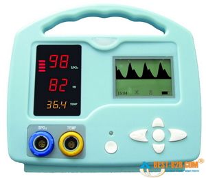
Most oximeters give you a few different pieces of information when they’re applied. The most important is the SaO2, a percentage between 0% and 100% describing how saturated the hemoglobin are with oxygen. (Typically, in most cases we refer to this number as SpO2, which is simply SaO2 as determined by pulse oximetry. This can be helpful by reminding us that oximeters aren’t perfect, and aren’t necessarily giving us a direct look at the blood contents, but for most purposes they are interchangeable terms.) But due to the pulse detection we just described, most oximeters will also display a fairly reliable heart rate for you.

Small handheld oximeters stop there. But larger models, such as the multi-purpose patient monitors used by medics and at hospital bedsides, will also display a waveform. This is a graphical display of the pulsatile flow, with time plotted on the horizontal axis and strength of the detected pulse on the vertical. With a strong, regular pulse, this waveform should be clear and regular, usually with peaked, jagged, or saw-tooth waves. Very small irregular waves, or a waveform with a great deal of artifact, is an indicator that the oximeter is getting a weak signal, and the calculated SpO2 (as well as the calculated pulse) may not be accurate. This waveform can also be used as a kind of “ghetto Doppler,” to help look for the presence of any pulsatile flow in extremities where pulses are not readily palpable. (To be technical, this waveform is known as a photoplethysmograph, or “pleth” for short, and potentially has other applications too– but we’ll leave it alone for now.)

Most modern oximeters, properly functioning and calibrated, have an accuracy between 1% and 2% — call it 1.5% on average. However, their accuracy falls as the saturation falls, and it is generally felt that at saturations below 70% or so, the oximeter ceases to provide reliable readings. Since sats below 90% or so correspond to the “steep” portion of the oxyhemoglobin dissociation curve, where small PaO2 changes might correspond to large changes in SpO2 — in other words, an alarming change in oxygenation status — the fact that your oximeter is losing accuracy in the ranges where you most rely on it is something to keep in mind if using oximetry for continuous monitoring.

The lag time between a change in respiratory conditions (such as increasing supplemental O2 or changing the ventilatory rate) and fully registering this change on the oximeter is usually around 1 minute. And at any given time, the displayed SpO2 is a value calculated by averaging the signal over several seconds, so any near-instantaneous changes should be considered false readings.

Keep reading for our next installment, when we discuss the clinical application of oximetry, and understanding false readings.

The Rapid Initial Assessment: Look, Talk, Feel

The initial assessment (known to old-timers as the “primary survey,” but it’s all the same idea) is the first phase of patient contact. It’s the initial period where you aim your eyeballs at the human being you’re going to be caring for and uncover the most basic facts about them.

Nowadays it’s taught as a discrete series of steps, usually something like this:

  1. General impression
  2. Assess responsiveness: AVPU
  3. Assess life threats: ABCs
    1. Assess and manage airway
    2. Assess and support breathing
    3. Assess and support circulation
  4. Determine patient priority

All good stuff, and there’s a reason it’s taught this way. All of these steps are important, and in order to teach (and test) them, they have to be broken down and explicitly described.

But this can be a shame, because in reality, the initial assessment isn’t like a recipe for a cake — mix this, then add that, then stir, then bake. It’s a brief burst of information, compacted into a dense flash of simultaneous sight, sound, and touch, and it can always be completed within a few seconds. In many cases it will be near instantaneous. In some it might take up to ten seconds. But it should never take as long as you’d need to actually verbalize all the steps.

The initial assessment should be a tight, elegant performance, and it’s one of the EMT’s most important skills. In the field, patients don’t come with charts or reports; all we know is what we’re dispatched with, which is usually wrong. But 90% of what you need to know about the patient can be learned promptly in the initial assessment. This is how you orient yourself to the situation and discover immediate life threats; more information and a more detailed assessment will follow, and it may reveal important findings, but our most critical job is to discover and treat what’s killing them, and that happens in the initial assessment. If you never got past this step you’d still be doing all of the most important things for the sickest people.

Here’s the process I recommend. It condenses everything you need to know into three simple steps.


Step 1: Look

You walk up and encounter your patient. What do you see?

Is he standing? Then he’s certainly conscious and alert. Is he moving purposefully or talking? Same business. Is he lying on the ground unconscious? We’ll learn more in a moment.

If he’s talking, his airway is intact and likely secure. You can roughly assess his breathing in about two seconds. Is he gasping for breath? Is he apneic? Is he speaking in full sentences?

Look at his skin. Is it pink? Is it pale and sweaty? Is it cyanotic? Is there obvious major trauma, such as significant bleeding anywhere or a puncture wound to the chest?


Step 2: Talk

Greet the patient and introduce yourself. “Hi, I’m Brandon.”

On a 911 response, you then ask for the patient’s name. How does he respond? Does he fail to recognize your presence at all? Does he look at you, but say nothing? Does he respond with a moan? Does he respond with, “George,” but his wife shakes her head and tells you otherwise? Does he promptly tell you his name?

To hear your words and verbalize an appropriate response requires alertness, engagement, memory, eye movement, vocal activity, and more. It requires the use of his airway and respiratory system, and thus reveals much about their status. Is he gurgling as he breathes? Gasping? You’ve learned a great deal already.

If you’re transferring a patient from a facility, you will already know the patient’s name, and pretending otherwise may make them wonder if you’ve got the wrong room. Better to skip their name and ask instead how they’re feeling. This leads you right into their chief complaint and subjective wellness, which is another huge slice of information. Are they in pain? Nauseous? Dizzy?


Step 3: Touch

As you talk, grasp the patient’s arm. You might politely interject, “May I grab you?” as appropriate.

Feel his skin. Is it dry, moist, or wet? Is it warm, hot, cool, or cold?

Feel his radial pulse. Is it present or absent? Is it weak, strong, or bounding? Is it slow or rapid, regular or irregular? There’s no need to count; that can wait for a full, proper set of vitals, which will come after our initial assessment. We’re just looking for a quick snapshot here.

This single touch tells you all sorts of things about his circulatory status. A patient with warm skin and a strong, regular radial pulse almost certainly has adequate volume and no immediate systemic crises. And anyway, taking someone by the hand is comforting in a primal way.

Let’s watch a few examples of this process at work.


Dispatched: MVA

Upon your arrival, you see a sedan in the middle of the road, with minor damage to the front bumper and right quarter panel. Beside it, you see an adult male walking around, slightly obese but appearing generally well.

He is ambulating easily and has no obvious bleeding or deformities. He therefore has a patent airway, largely adequate breathing and circulation, and his general impression is good. You could stop here, but we won’t.

You approach him, saying with a smile, “Hi, I’m Brandon. What’s your name?” He replies, “Greg Rogers — some idiot tried to pull out in front of me.” His breathing appears unlabored. As you talk, you take him by the wrist, feeling warm, dry skin and a strong, regular, slightly rapid radial pulse.

He appears neurologically intact, with good memory and appropriate responses. His breathing is normal and his circulation appears fine, although he is obviously a little excited.

[Initial asessment complete. Total time: 1 second to learn everything important; 5 seconds from soup to nuts. He has no life threats and is a low transport priority.]


Dispatched: Welfare check

You walk in the room to find an elderly woman supine on the bed, curled in an awkward position and motionless.

You are already highly suspicious of a depressed level of consciousness. It is possible she is merely sleeping, but most people would not sleep in such a position.

Approaching, you lean over and call, “Ma’am! Can you hear me?!” You gently shake her shoulder while you do. There is no response.

She is not alert. This is the “are you napping?” test; if she were easily roused in the same way you’d wake up your roommate, we would call her alert, not “responsive to voice”. You don’t lose points just for being asleep.

You lean into her ear and call again, this time in a loud shout. There is no response.

She is unresponsive to verbal stimuli. A loud, intrusive sound elicited no reaction.

Rolling her over, you note the sound of snoring respirations. Her chest is rising and falling with good depth, but not very quickly. Her skin is slightly ashen. You give her brachial plexus a tight pinch, to which she flinches and withdraws slightly.

She is responsive to painful stimuli, but does not open her eyes. (If you later wanted to calculate her GCS, she would earn a 5.) Her airway needs managing, and an OPA would probably be appropriate. She should receive supplemental oxygen as well, and may require assistance with the BVM. Since she’s breathing, she presumably has a pulse.

With one hand, you palpate her carotid pulse, while you palpate her radial pulse with the other. Her pulses are regular and slightly slow. Her radial is strong, and her skin is warm and dry both at the neck and at the wrist.

She has adequate circulation, perhaps with a slight bradycardia due to hypoxia. Her volume is adequate.

[Initial assessment complete. Total time: 6 seconds. She will need airway and breathing support, then a rapid assessment and transport due to her diminished level of consciousness.


Dispatched: Discharge to skilled nursing

You walk into the hospital room to find your patient in bed, semi-Fowler’s. Her eyes are open and staring at the ceiling, but she makes no acknowledgement of your presence. She is breathing adequately and without labor. Her skin appears dry and slightly pale.

She appears conscious, has an airway, and is breathing. She presumably has a pulse. She appears unremarkable for an ill but stable elderly patient, perhaps with a baseline dementia.

You approach her, saying, “Ms. Smith!” She turns her head and makes eye contact. “I’m Brandon. How are you feeling?” She replies, “Hi…” After another couple attempts, the best response she gives is to call you “Aaron” and ask about the elephants.

She is alert and engaged with her surroundings, but poorly oriented and disconnected with reality.

While you talk, you ask if you can see her arm; she pulls it slightly out from the sheets. You take her wrist with one hand. Her skin is pale, dry, and slightly cool peripherally, with poor turgor. Her radial pulse is very weak and irregularly irregular.

She is able to follow commands, but physically weak. Her peripheral circulation is poor, likely secondary to both poor cardiac output (her irregular pulse is consistent with atrial fibrillation) and peripheral vascular disease.

[Initial assessment complete. Total time: 8 seconds. Her presentation is consistent with her documented history and she is likely ready for transport.]

You may notice in all this that we haven’t performed any interventions — not even a lowly nasal cannula. The initial assessment is usually taught in a “treat as you assess” fashion; if you check the airway and find it compromised, you should address it before moving on. But look how fast we moved through all this! Wouldn’t you rather bang out your initial assessment in a few seconds, then move on to your treatments having a full knowledge of the situation? If we check the airway, and go to the trouble of sizing and inserting an OPA, by the time we’re done we still have no idea about breathing or circulatory status — something that would have taken another second or two to assess at most.

Initial assessments are like a flash of lightning: you start with nothing, and with a sudden burst of light, you end up with a great deal. That flash won’t tell you the whole story, and you’ll always need to keep looking and keep digging. But with a smart and efficient initial assessment, you’ll set the stage and choose the course for everything else to come. All in under ten seconds.

Get Up, Stand Up: Orthostatics

Orthostatic vital signs. Nurses think they’re a pain in the neck. Some doctors think they’re of marginal usefulness. Many providers simply think they’re a dying breed.

Like many old-school physical exam techniques, though, they’re dying only because high-tech imaging and laboratory techniques have largely replaced their role. And I don’t know about you, but my ambulance doesn’t come equipped for an ultrasound or serum electrolytes. Diagnostically, EMS lives in the Olden Days — the days of the hands-on physical, the stethoscope, the palpation and percussion, the careful and detailed history. For us, orthostatics have been and still are a valuable tool in patient assessment.

How are they performed? Orthostatic vital signs are essentially multiple sets of vitals taken from the patient in different positions. (They’re also sometimes known as the tilt test or tilt table, which is indeed another way to perform them — if you have a big, pivoting table available. Postural vitals is yet another name.) They usually include blood pressure and pulse, and are taken in two to three positions — supine (flat on the back) and standing are the most common, but a sitting position is sometimes also included, or used instead of standing. This is useful when a patient is unable to safely stand, although it’s not quite as diagnostically sensitive.

Why would we do such a dance? The main badness that orthostatics reveal is hypovolemia. With a full tank of blood, what ordinarily happens when I stand up? Gravity draws some of my blood into the lower portion of my body (mostly these big ol’ legs). This reduces perfusion to the important organs upstairs, especially my brain, so my body instantly compensates by increasing my heartrate a bit and tightening up my vasculature. No problem. However, what if my circulating volume is low — whether due to bleeding, dehydration, or even a “relative” hypovolemia (in distributive shocks such as sepsis or anaphylaxis)? In that case, when my smaller volume of blood is pulled away by gravity, my body will have a harder time compensating. If it’s not fully able to, then my blood pressure will drop systemically.

“But,” you cry, “surely this is all just extra steps. Can’t I recognize hypovolemia from basic vital signs — no matter what position you’re in?”

Well, yes and no. If it’s severe enough, then it will be readily apparent even if I’m standing on my head. But we routinely take baseline vitals on patients who are at least somewhat horizontal, and this is the ideal position to allow the body to compensate for low volume. By “challenging” the system with the use of gravity, we reveal the compensated hypovolemias… rather than only seeing the severely decompensated shock patients, who we can easily diagnose from thirty paces anyway. Like a cardiac stress test, we see more by pushing the body until it starts to fail; that’s how you discover the cracks beneath the surface.

Do we run on patients with hypovolemia? Oh, yes. External bleeding is a gimme, but how about GI bleeds? Decreased oral fluid intake? Increased urination due to diuretics? How about the day after a frat party kegger? Any of this sound familiar? It would be foolish to take the time to do this when it won’t affect patient care — such as in the obviously shocked patient — but there are times when what it reveals can be important, such as in patients who initially appear well and are considering refusing transport.

Here’s the process I’d recommend for taking orthostatics:

  1. Start with your initial, baseline set of vitals. Whatever position your patient is found in, that’s fine. Deal with your initial assessment in the usual fashion.
  2. Once you’re starting to go down a diagnostic pathway that prominently includes hypovolemic conditions in the differential, start thinking about orthostatics. If your initial vitals were taken while seated, try lying the patient flat and taking another pulse and BP. If possible, wait a minute or so between posture change and obtaining vitals; this will allow their system to “settle out” and avoid capturing aberrant numbers while they reestablish equilibrium.
  3. Ask yourself: can the patient safely stand? Even in altered or poorly-ambulatory individuals, the answer might be “yes” with your assistance, up to and including a burly firefighter supporting them from behind with a bearhug. (Caution here is advised even in basically well patients, because significant orthostatic hypotension may result in a sudden loss of consciousness upon standing. You don’t want your “positive” finding to come from a downed patient with a fresh hip fracture.) If safe to do so, stand the patient and take another pulse and BP. Again, waiting at least a minute is ideal, but if that’s not possible, don’t fret too much.
  4. For totally non-ambulatory patients, substitute sitting upright for standing. Ideally, this should be in a chair (or off the side of the stretcher) where their legs can hang, rather than a Fowler’s position with legs straight ahead.
  5. For utterly immobile patients who can’t even sit upright, or if attempting orthostatics in the truck while already transporting, you’ll need to do your best to position them with the stretcher back itself. Fully supine will be your low position, full upright Fowler’s will be your high position, and a semi-Fowler’s middle ground can be included if desired.

On interpretation: healthy, euvolemic patients can exhibit small orthostatic changes, so hypovolemia is only appreciable from a significant drop in BP or increase in heart rate. From supine to standing, a drop in the systolic blood pressure of over 20 is usually considered abnormal, as is an increase in pulse of over 30. (Changes from supine to sitting, or sitting to standing, will obviously be smaller, and therefore harder to distinguish from ordinary physiological fluctuations.) A drop in diastolic pressure of over 10 is also considered aberrant. You can remember this as the “10–20–30” rule.

Try to remember what’s going on here. As the patient shifts upright, their available volume is decreasing, for which their body attempts to compensate — in part by increasing their heart rate. It’s a truism that younger, healthier, less medicated patients are more able to compensate than older and less well individuals. So for the same volume status, you would be more likely to see an increase in pulse from the younger patient, perhaps with no change in pressure; whereas the older patient might have less pulse differential but a greater drop in pressure. (On the whole, the pulse change tends to be a more sensitive indicator than pressure, since almost everyone is able to compensate somewhat for orthostatic effects. As always, if you look for the compensation rather than the decompensation — the patch, rather than the hole it’s covering — you’ll see more red flags and find them sooner.)

Are substantial orthostatic changes definitive proof of hypovolemia? No, nothing’s certain in this world. Another possible cause is autonomic dysregulation, which essentially means that the normal compensating mechanisms (namely baroreceptors that detect the drop in pressure and stimulate vasoconstriction, chronotropy, and inotropy) fail to function properly. You do have enough juice, but your body isn’t doing its job of keeping it evenly circulating. Vasovagal syncope is one common example of this; I’ve got it myself, in fact, and hence have a habit of passing out while squatting. This sort of thing is not related to volume status, although if you combine the two the effect can be synergistic. A good history can help distinguish them: ask the patient if they have a prior history of dizziness upon standing.

Finally, pulse and pressure are not the only changes you can assess. One of the best indicators of orthostatic hypotension is simply a subjective feeling of light-headedness reported by the patient. Although sudden light-headedness upon standing can have other causes (the other big possibility is benign paroxysmal positional vertigo — although strictly speaking, BPPV tends to cause “dizziness,” which is not the same as “lightheadedness”), hypovolemia is certainly one of the most likely. So stand ’em up when it’s safe and reasonable, ask how they feel, grab the vitals if you can, and maybe even take the opportunity to see how well they walk (a nice, broad neurological test — the total inability to ambulate in a normally ambulatory patient is a very ominous sign).

Orthostatics are usually recorded on documentation by drawing little stick figures of the appropriate postures. For those who find this goofy, or are documenting on computers without “stick figure” keys, a full written description will do.

The Rhythm Method


One two three — five six seven

What’s the missing number?

If you said four, congratulations. You have a basic human ability to recognize patterns — one of the best tools we have to separate us from the monkeys and sea-slugs.

One of the simplest types of pattern is a rhythm, and the simplest rhythm is a steady cadence. Ba-dump, ba-dump, ba-dump. Imagine a metronome or a drummer tapping out a fixed, continuous pace at an unchanging rhythm.

This is also one of the most basic and useful tricks you’ll ever use when taking vitals!

See, measuring vitals involves feeling, hearing, or observing a series of fairly subtle blips over a period of time. Unfortunately, interference is common in the field, and it’s a rare day when bumps in the road and bangs in the cabin don’t eat up at least one of those blips.

When taking a radial pulse, if over 15 seconds you count 18 beats, you have a pulse of 72; but if just a couple of those beats are lost due to your movement or the patient’s, suddenly it becomes 64, which is a substantial difference. This is no good; we want better reliability than that.

Rhythm is the answer. A pulse is typically a regular rhythm. So are respirations. So are the Korotkoff sounds of a blood pressure. In order to establish this rhythm, you only need to hear two consecutive beats, and appreciate exactly how far apart they are. If you can do this, then you can continue to mentally tap out that pace — hopefully, while continuing to feel, see, or hear the true beats, which will help you to maintain the right speed, but even if you miss some, you’ll still have your mental beat to count. Even if you miss most of them!

So you feel for the pulse, and you palpate the first couple beats. Then you hit a tortuous section of road that throws you around the cabin, and you’re unable to feel anything for several seconds. But you already had the rhythm in your head, so when you pick up the pulse again, you haven’t lost the count — and you’ll end up with an accurate number.

Now, in sick people these rhythms aren’t always regular. And if you observe that a pulse or respiratory cycle isn’t regular, then this system won’t be as effective — for instance, there’s not much point in trying to find the “beat” to an A-Fib pulse. But small irregularities or breaks in the rhythm are okay, as long as there’s still a regular cycle underlying it; for instance, occasional dropped (or extra) beats won’t change the basic rate.

Give it a try. If you got rhythm, vital signs will never give you trouble again.

Vital Signs: Pulse

For other Vital Signs posts, see: Respirations and Blood Pressure

Ah, the almighty pulse. If I have a favorite vital sign, this is it; let me lay hands on a patient and take a pulse and my assessment is already well under way.

On the conscious patient our go-to point is the radial pulse, and like golf, mastering the radial is all in the grip. Techniques may vary here, but I always find the radial easier to palpate if you approach from the ulnar side of the arm, coming “underneath” rather than over the top of the radius. This also lets you take a pulse while easily holding onto their limb, rather than forcing you to find a place to rest it, or supporting the arm with one hand while you palpate with the other. Just grab and count, very natural. If you have no luck, you can always keep hold of their arm while using your other hand to do some searching.

The textbooks always seem to show this being done with two delicate fingers, which is silly; more fingers means more coverage, so I always use at least three. (Your little finger is kinda short, otherwise it’d be four.) Use a moderate pressure, but if you’re having trouble, try pressing both lighter and firmer, as well as moving to different spots. (While I usually wear my watch in the normal position, you’ll notice here that when taking a pulse this way, I flip it around my wrist so I can see the face.)

The main way to ensure you’re never baffled by the pulse, however, is by always being willing to look elsewhere. Some people simply won’t have a radial, and this fact may or may not have significance — it may mean they’re hypotensive, or that their arm is locally hypoperfused, but it also may be a chronic condition. Hemodialysis patients with arterio-venous fistulas in their arm are especially notorious for having peculiar or absent radial pulses, as the arteries near the fistula have been scavenged and rerouted. Make like a picky renter — go elsewhere!

Your next attempt after the radial should be the brachial. Now, in classes and textbooks I have always been taught to look for a radial in the upper arm, beneath the bicep, but I’ve never had luck with this. Rather, my target is the antecubital fossa, the same territory made popular by blood pressures and large-bore IV sticks.

Again, positioning is key here. To effectively feel this pulse, the elbow should be in full extension, but relaxed. Depending on the patient’s position, you may accomplish this by wrapping your arm around theirs and holding their elbow in your hand, but from your bench seat in the truck, an easier way to do it is to simply rest their elbow on your knee. (Either way, it’s important to support them at the elbow, because this allows gravity to force their arm into extension.) The brachial can be a real lifesaver when a radial isn’t forthcoming, and I go to it readily and often.

Logically, the next step would be a carotid pulse, but the truth is that on conscious, alert patients, this is always a little awkward; people don’t like having their neck touched. If they need it, they need it, but for the routine pulse check, I try to steer clear. The same goes for a femoral pulse, for the same reasons; there was a story at my old service of a brash young EMT who got canned for “feeling a femoral” on an inebriated coed from a campus we served.

Instead, if I can’t find a radial or brachial on either arm, I’ll often take an apical “pulse,” simply auscultating at the chest for heart sounds. This is not, strictly speaking, a pulse, insofar as it’s not counting actual perfusing beats so much as counting any cardiac noise (it therefore tells you nothing about blood pressure), but it’s a good fallback — and if you’re very suave it can even yield additional clinical information, regarding murmurs, rubs, etc.

Here are a some other tricks that can be useful:

  • Inflate a BP cuff and count the bounces on the sphygmomanometer needle. Although this is not an indicator of systolic or diastolic pressure, it is a legitimate way to measure a pulse.
  • If pulse oximetry is available, the device will usually calculate a pulse for you, and if there’s a displayed waveform you can also confirm it from that.
  • The aforementioned AV fistulas can be used to your advantage. Gentle palpation of visible, active fistulas should let you feel a pulsing vibration called a thrill (an indicator of healthy flow), and this is easily counted for an accurate pulse rate. (Auscultating at the fistula should reveal a buzzing sound called bruit, which can be used similarly.)
  • If you’re able to locate a difficult pulse point, such as a dorsalis pedis, X’ng the spot with a pen can make subsequent checks much easier.
  • Lowering the arm below the level of the heart can occasionally make a radial more readily palpable, especially in hypotensive situations.

Finally, when all else fails, remember your perpetual fallback: skin signs. A patient with no available pulses and no obtainable blood pressure can still give you a general sense of perfusion, both centrally and to each extremity, if you assess the color and temperature of his skin. (This is especially valuable for infants, for whom proper pulse checks can be difficult, and blood pressures even more so.) And then there’s the sidekick to this, which is capillary refill. Current teaching is that cap refill is not a meaningful sign except in the very young, because numerous chronic conditions can cause delayed refill without poor arterial pressure, and this is true; a slow cap refill in an adult shouldn’t mean much to you. However, a rapid refill is still a pretty specific sign of good perfusion, because there’s not many conditions that can fake that (with the possibly exception of distributive shocks, such as septic or anaphylactic). A quick pat-down is an ever-ready way to rapidly assess anyone’s hemodynamic status within a couple seconds.