Get Up, Stand Up: Orthostatics

Orthostatic vital signs. Nurses think they’re a pain in the neck. Some doctors think they’re of marginal usefulness. Many providers simply think they’re a dying breed.

Like many old-school physical exam techniques, though, they’re dying only because high-tech imaging and laboratory techniques have largely replaced their role. And I don’t know about you, but my ambulance doesn’t come equipped for an ultrasound or serum electrolytes. Diagnostically, EMS lives in the Olden Days — the days of the hands-on physical, the stethoscope, the palpation and percussion, the careful and detailed history. For us, orthostatics have been and still are a valuable tool in patient assessment.

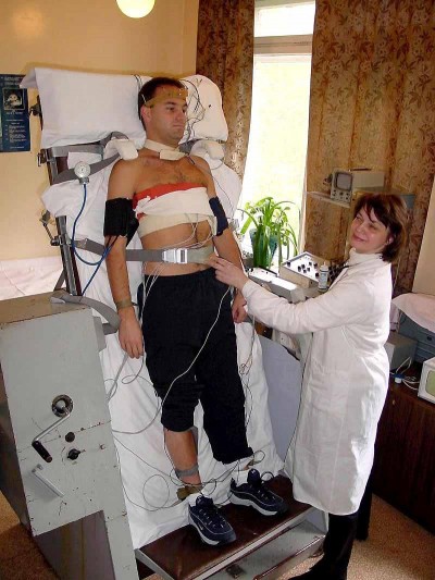
How are they performed? Orthostatic vital signs are essentially multiple sets of vitals taken from the patient in different positions. (They’re also sometimes known as the tilt test or tilt table, which is indeed another way to perform them — if you have a big, pivoting table available. Postural vitals is yet another name.) They usually include blood pressure and pulse, and are taken in two to three positions — supine (flat on the back) and standing are the most common, but a sitting position is sometimes also included, or used instead of standing. This is useful when a patient is unable to safely stand, although it’s not quite as diagnostically sensitive.

Why would we do such a dance? The main badness that orthostatics reveal is hypovolemia. With a full tank of blood, what ordinarily happens when I stand up? Gravity draws some of my blood into the lower portion of my body (mostly these big ol’ legs). This reduces perfusion to the important organs upstairs, especially my brain, so my body instantly compensates by increasing my heartrate a bit and tightening up my vasculature. No problem. However, what if my circulating volume is low — whether due to bleeding, dehydration, or even a “relative” hypovolemia (in distributive shocks such as sepsis or anaphylaxis)? In that case, when my smaller volume of blood is pulled away by gravity, my body will have a harder time compensating. If it’s not fully able to, then my blood pressure will drop systemically.

“But,” you cry, “surely this is all just extra steps. Can’t I recognize hypovolemia from basic vital signs — no matter what position you’re in?”

Well, yes and no. If it’s severe enough, then it will be readily apparent even if I’m standing on my head. But we routinely take baseline vitals on patients who are at least somewhat horizontal, and this is the ideal position to allow the body to compensate for low volume. By “challenging” the system with the use of gravity, we reveal the compensated hypovolemias… rather than only seeing the severely decompensated shock patients, who we can easily diagnose from thirty paces anyway. Like a cardiac stress test, we see more by pushing the body until it starts to fail; that’s how you discover the cracks beneath the surface.

Do we run on patients with hypovolemia? Oh, yes. External bleeding is a gimme, but how about GI bleeds? Decreased oral fluid intake? Increased urination due to diuretics? How about the day after a frat party kegger? Any of this sound familiar? It would be foolish to take the time to do this when it won’t affect patient care — such as in the obviously shocked patient — but there are times when what it reveals can be important, such as in patients who initially appear well and are considering refusing transport.

Here’s the process I’d recommend for taking orthostatics:

  1. Start with your initial, baseline set of vitals. Whatever position your patient is found in, that’s fine. Deal with your initial assessment in the usual fashion.
  2. Once you’re starting to go down a diagnostic pathway that prominently includes hypovolemic conditions in the differential, start thinking about orthostatics. If your initial vitals were taken while seated, try lying the patient flat and taking another pulse and BP. If possible, wait a minute or so between posture change and obtaining vitals; this will allow their system to “settle out” and avoid capturing aberrant numbers while they reestablish equilibrium.
  3. Ask yourself: can the patient safely stand? Even in altered or poorly-ambulatory individuals, the answer might be “yes” with your assistance, up to and including a burly firefighter supporting them from behind with a bearhug. (Caution here is advised even in basically well patients, because significant orthostatic hypotension may result in a sudden loss of consciousness upon standing. You don’t want your “positive” finding to come from a downed patient with a fresh hip fracture.) If safe to do so, stand the patient and take another pulse and BP. Again, waiting at least a minute is ideal, but if that’s not possible, don’t fret too much.
  4. For totally non-ambulatory patients, substitute sitting upright for standing. Ideally, this should be in a chair (or off the side of the stretcher) where their legs can hang, rather than a Fowler’s position with legs straight ahead.
  5. For utterly immobile patients who can’t even sit upright, or if attempting orthostatics in the truck while already transporting, you’ll need to do your best to position them with the stretcher back itself. Fully supine will be your low position, full upright Fowler’s will be your high position, and a semi-Fowler’s middle ground can be included if desired.

On interpretation: healthy, euvolemic patients can exhibit small orthostatic changes, so hypovolemia is only appreciable from a significant drop in BP or increase in heart rate. From supine to standing, a drop in the systolic blood pressure of over 20 is usually considered abnormal, as is an increase in pulse of over 30. (Changes from supine to sitting, or sitting to standing, will obviously be smaller, and therefore harder to distinguish from ordinary physiological fluctuations.) A drop in diastolic pressure of over 10 is also considered aberrant. You can remember this as the “10–20–30” rule.

Try to remember what’s going on here. As the patient shifts upright, their available volume is decreasing, for which their body attempts to compensate — in part by increasing their heart rate. It’s a truism that younger, healthier, less medicated patients are more able to compensate than older and less well individuals. So for the same volume status, you would be more likely to see an increase in pulse from the younger patient, perhaps with no change in pressure; whereas the older patient might have less pulse differential but a greater drop in pressure. (On the whole, the pulse change tends to be a more sensitive indicator than pressure, since almost everyone is able to compensate somewhat for orthostatic effects. As always, if you look for the compensation rather than the decompensation — the patch, rather than the hole it’s covering — you’ll see more red flags and find them sooner.)

Are substantial orthostatic changes definitive proof of hypovolemia? No, nothing’s certain in this world. Another possible cause is autonomic dysregulation, which essentially means that the normal compensating mechanisms (namely baroreceptors that detect the drop in pressure and stimulate vasoconstriction, chronotropy, and inotropy) fail to function properly. You do have enough juice, but your body isn’t doing its job of keeping it evenly circulating. Vasovagal syncope is one common example of this; I’ve got it myself, in fact, and hence have a habit of passing out while squatting. This sort of thing is not related to volume status, although if you combine the two the effect can be synergistic. A good history can help distinguish them: ask the patient if they have a prior history of dizziness upon standing.

Finally, pulse and pressure are not the only changes you can assess. One of the best indicators of orthostatic hypotension is simply a subjective feeling of light-headedness reported by the patient. Although sudden light-headedness upon standing can have other causes (the other big possibility is benign paroxysmal positional vertigo — although strictly speaking, BPPV tends to cause “dizziness,” which is not the same as “lightheadedness”), hypovolemia is certainly one of the most likely. So stand ’em up when it’s safe and reasonable, ask how they feel, grab the vitals if you can, and maybe even take the opportunity to see how well they walk (a nice, broad neurological test — the total inability to ambulate in a normally ambulatory patient is a very ominous sign).

Orthostatics are usually recorded on documentation by drawing little stick figures of the appropriate postures. For those who find this goofy, or are documenting on computers without “stick figure” keys, a full written description will do.

The Rhythm Method


One two three — five six seven

What’s the missing number?

If you said four, congratulations. You have a basic human ability to recognize patterns — one of the best tools we have to separate us from the monkeys and sea-slugs.

One of the simplest types of pattern is a rhythm, and the simplest rhythm is a steady cadence. Ba-dump, ba-dump, ba-dump. Imagine a metronome or a drummer tapping out a fixed, continuous pace at an unchanging rhythm.

This is also one of the most basic and useful tricks you’ll ever use when taking vitals!

See, measuring vitals involves feeling, hearing, or observing a series of fairly subtle blips over a period of time. Unfortunately, interference is common in the field, and it’s a rare day when bumps in the road and bangs in the cabin don’t eat up at least one of those blips.

When taking a radial pulse, if over 15 seconds you count 18 beats, you have a pulse of 72; but if just a couple of those beats are lost due to your movement or the patient’s, suddenly it becomes 64, which is a substantial difference. This is no good; we want better reliability than that.

Rhythm is the answer. A pulse is typically a regular rhythm. So are respirations. So are the Korotkoff sounds of a blood pressure. In order to establish this rhythm, you only need to hear two consecutive beats, and appreciate exactly how far apart they are. If you can do this, then you can continue to mentally tap out that pace — hopefully, while continuing to feel, see, or hear the true beats, which will help you to maintain the right speed, but even if you miss some, you’ll still have your mental beat to count. Even if you miss most of them!

So you feel for the pulse, and you palpate the first couple beats. Then you hit a tortuous section of road that throws you around the cabin, and you’re unable to feel anything for several seconds. But you already had the rhythm in your head, so when you pick up the pulse again, you haven’t lost the count — and you’ll end up with an accurate number.

Now, in sick people these rhythms aren’t always regular. And if you observe that a pulse or respiratory cycle isn’t regular, then this system won’t be as effective — for instance, there’s not much point in trying to find the “beat” to an A-Fib pulse. But small irregularities or breaks in the rhythm are okay, as long as there’s still a regular cycle underlying it; for instance, occasional dropped (or extra) beats won’t change the basic rate.

Give it a try. If you got rhythm, vital signs will never give you trouble again.

What it Looks Like: Agonal Respirations

See also what Jugular Venous DistentionSeizures, and Cardiac Arrest and CPR look like

Education and experience are both important to making a well-rounded provider, and each of the two have distinct advantages. Perhaps the greatest advantage of experience is that it gives you the best ability to recognize situations you’d otherwise only know by description or by photograph.

Nowadays, though, with the Wonders of Modern Technology, we have some tools that can help bridge this gap. Experience is still essential — but there’s no reason that the first time you see a seizure or cyanosis should be in a situation with real stakes.

So let’s go through some of the common medical events and conditions we talk about, learn about, but may not truly know the presentation of until we encounter it.

Today, it’s:


Agonal Respirations

Agonal respirations are an inadequate pattern of breathing associated with extreme physiological distress, particularly periarrest states (that is, it is usually seen just prior to cardiac arrest, as well as during and for some time after). Although not always seen during arrest, it is not uncommon, and there is some evidence that it may be associated with better outcomes than arrests without agonal breathing. Whatever the case, it can easily be confused for ordinary respiration, leading to the mistaken impression that the “breathing” patient must also have a pulse; this confusion is part of why the American Heart Association no longer recommends checking for breathing as part of layperson’s CPR.

As for healthcare providers, whether we’re able to put the label of “agonal” on it or not, we should be able to recognize from the rate and depth that this is not adequate respiration to sustain oxygenation, and ventilatory assistance (as well as a check of hemodynamic status) is in order. But recognizing the specific nature of this breathing can be a very useful red flag to set your “code” wheels in motion.

Here are a few simulated examples, performed by medical actors. They range in presentation and context.

Finally, here’s a treat — this is a video of a real-life cardiac arrest at a beach in Australia. Starting after the first shock, from 2:39 onward, you can see a great example of agonal breathing. The rest of the video is also a nice example of an honest code being worked in the field — not perfect, but real. (For bonus points, how could their CPR and other treatment have been improved?)

(Thanks to Dave Hiltz for inspiring today’s topic.)

Vital Signs: Blood Pressure

For other Vital Signs posts, see: Respirations and Pulse

In the grand scheme of medical skills, taking a manual blood pressure is far from difficult, but sick people and austere conditions can combine to make it another thing entirely. Obtaining a BP on an ill patient while rattling down the road is legitimately one of the most difficult psychomotor skills an EMT-Basic has to master.

Mastering it starts with stacking the odds in your favor. A good stethoscope is better than a lousy one — you don’t need a $500 cardiology model, but something with good insulation and tight-fitting earpieces can make a real difference. Of course, you’ll also want to try to take your blood pressures at times of peace: on scene, before the rig starts moving, or even shoehorned in while stopped at traffic lights.

The elbow-supported technique for finding the brachial pulse is also ideal for taking a BP; trying to hear anything when the arm is slightly flexed is a recipe for frustration. But ensure that however you arrange things, the arm is completely relaxed, because muscular tension can radically throw a measurement; this will require fully supporting the arm and sometimes reassuring the patient. “Just relax” is the line I always deliver while busily pumping the bulb.

Where to put the gauge? Wherever. I’ll usually clip it to one of the stretcher straps, but you can find a bit of blanket that it’ll nestle into, secure it to a shirt, clip it to your watchband or the edge of the cuff, or just ask the patient to hold it for you. The built-in strap on the cuff is only a good location if you’re at the patient’s right side, which is typically not where we sit while we’re transporting. There’s probably a huge market niche out there for “EMS style” cuffs with their handedness reversed… but I digress.

Although I don’t always follow all of these steps, here’s the basic approach I recommend for a routine blood pressure check:

  • Support the arm, ideally at a position that is horizontally level with the heart.
  • Palpate the antecubital fossa until you find the pulse point. Note this location.
  • Palpating at the radial or the AC, pump up the cuff until you lose the pulse. Note this number and deflate the cuff.
  • Place your scope on the AC and inflate the cuff past the previous number. Obtain your pressure in the ordinary fashion.

Starting with a palpated pressure may seem redundant, and it can be, but it has two advantages: first, it gives you a rough sense of what systolic to look for, and second, if you’re unable to auscultate a pressure, you’ll still have a palpated one to record. This is actually the officially recommended method, although it seems rarely done nowadays.

Palpated pressures are legitimate, although when they start becoming the norm it can be a sign of lazy care. The diastolic can be a valuable number, though, particularly in traumatic or cardiac cases, so remember that auscultating is still the default standard of care. And remember, particularly if you’re mixing methods, that palpated pressures often will differ from auscultated pressures (including those taken by machine), usually by 10-15 points on the low side.

What if you’re not getting anything from the arm? Well, you can try the other arm, of course. But really, the thing to remember is that you can take a blood pressure anywhere there’s a pulse, although it’s much easier when that pulse is strong and the artery proximal to it can be easily occluded. Remember that although you can palpate a pressure from any distal spot on the same artery, near or far (barring anastamoses), auscultation — which is essentially listening to the turbulence created immediately downstream of the occlusion — requires placing your scope just below the cuff, and will not be successful farther downstream. Putting the cuff (pedi cuffs when needed) on the forearm and measuring at the radial is effective; thigh cuffs work too, although the popliteal can be an evasive pulse to locate. You can even cuff the lower calf and palpate a pedal or tibial pulse, if you’re daring. Go nuts, and try to experiment before the call when you actually need it. Do make an effort, though, to use an appropriate sized cuff for the extremity; mis-sized cuffs can actually yield significantly erroneous readings. For the morbidly obese, I usually prefer to place a regular cuff on the forearm than to use a thigh cuff on the upper arm, but see what works for you.

As a final note, remember that cuffing the neck and palpating the temporal pulse is never an appropriate method of patient assessment, no matter how little blood you may suspect is reaching their brain.

On maintenance: during your morning checkout, pump some air into the cuff, close the valve and give the whole thing a squeeze to check for leaks. There’s nothing better than discovering these after you’ve wrapped it around a critical patient’s arm.

On sphygmomanometers: for obvious reasons, the resting point for the needle should be at zero. (Very cheap cuffs sometimes have a pin-stop here for the needle to rest against; this is a problem because the dial can be miscalibrated without showing it. Pin-stop gauges shouldn’t be used unless your service is seriously broke.) If you have one that needs zeroing, most cuffs can be adjusted by pulling the tubing off the dial, grasping the metal nipple with some pliers (or very strong fingers), and twisting it in either direction until the needle is zeroed. Alternately, fans of mental math can just add or subtract the false “zero” number each time they take a pressure.

And finally, on tourniquets: the immortal Dr. Scott Weingart of Emcrit has described his practice of using BP cuffs as tourniquets. You’ll hear about this from time to time, but there’s always someone who points out the damned things leak like sieves and that’s the last property you want in a tourniquet. Dr. Weingart’s solution is to pump up the cuff until bleeding is controlled (or 250mmHg, whichever is sooner), then clamp both tubes with locking hemostats. (He uses smooth ones to avoid damaging the rubber; he recommends padding with a 4×4 if you’re using a ridged hemostat.) My hemostats are all in the shop, and this may or may not fly with your agency — modifying equipment for “off-label” use is always somewhat shaky ground for us field peons — but I think it’s a splendid idea if you can swing it.

Vital Signs: Pulse

For other Vital Signs posts, see: Respirations and Blood Pressure

Ah, the almighty pulse. If I have a favorite vital sign, this is it; let me lay hands on a patient and take a pulse and my assessment is already well under way.

On the conscious patient our go-to point is the radial pulse, and like golf, mastering the radial is all in the grip. Techniques may vary here, but I always find the radial easier to palpate if you approach from the ulnar side of the arm, coming “underneath” rather than over the top of the radius. This also lets you take a pulse while easily holding onto their limb, rather than forcing you to find a place to rest it, or supporting the arm with one hand while you palpate with the other. Just grab and count, very natural. If you have no luck, you can always keep hold of their arm while using your other hand to do some searching.

The textbooks always seem to show this being done with two delicate fingers, which is silly; more fingers means more coverage, so I always use at least three. (Your little finger is kinda short, otherwise it’d be four.) Use a moderate pressure, but if you’re having trouble, try pressing both lighter and firmer, as well as moving to different spots. (While I usually wear my watch in the normal position, you’ll notice here that when taking a pulse this way, I flip it around my wrist so I can see the face.)

The main way to ensure you’re never baffled by the pulse, however, is by always being willing to look elsewhere. Some people simply won’t have a radial, and this fact may or may not have significance — it may mean they’re hypotensive, or that their arm is locally hypoperfused, but it also may be a chronic condition. Hemodialysis patients with arterio-venous fistulas in their arm are especially notorious for having peculiar or absent radial pulses, as the arteries near the fistula have been scavenged and rerouted. Make like a picky renter — go elsewhere!

Your next attempt after the radial should be the brachial. Now, in classes and textbooks I have always been taught to look for a radial in the upper arm, beneath the bicep, but I’ve never had luck with this. Rather, my target is the antecubital fossa, the same territory made popular by blood pressures and large-bore IV sticks.

Again, positioning is key here. To effectively feel this pulse, the elbow should be in full extension, but relaxed. Depending on the patient’s position, you may accomplish this by wrapping your arm around theirs and holding their elbow in your hand, but from your bench seat in the truck, an easier way to do it is to simply rest their elbow on your knee. (Either way, it’s important to support them at the elbow, because this allows gravity to force their arm into extension.) The brachial can be a real lifesaver when a radial isn’t forthcoming, and I go to it readily and often.

Logically, the next step would be a carotid pulse, but the truth is that on conscious, alert patients, this is always a little awkward; people don’t like having their neck touched. If they need it, they need it, but for the routine pulse check, I try to steer clear. The same goes for a femoral pulse, for the same reasons; there was a story at my old service of a brash young EMT who got canned for “feeling a femoral” on an inebriated coed from a campus we served.

Instead, if I can’t find a radial or brachial on either arm, I’ll often take an apical “pulse,” simply auscultating at the chest for heart sounds. This is not, strictly speaking, a pulse, insofar as it’s not counting actual perfusing beats so much as counting any cardiac noise (it therefore tells you nothing about blood pressure), but it’s a good fallback — and if you’re very suave it can even yield additional clinical information, regarding murmurs, rubs, etc.

Here are a some other tricks that can be useful:

  • Inflate a BP cuff and count the bounces on the sphygmomanometer needle. Although this is not an indicator of systolic or diastolic pressure, it is a legitimate way to measure a pulse.
  • If pulse oximetry is available, the device will usually calculate a pulse for you, and if there’s a displayed waveform you can also confirm it from that.
  • The aforementioned AV fistulas can be used to your advantage. Gentle palpation of visible, active fistulas should let you feel a pulsing vibration called a thrill (an indicator of healthy flow), and this is easily counted for an accurate pulse rate. (Auscultating at the fistula should reveal a buzzing sound called bruit, which can be used similarly.)
  • If you’re able to locate a difficult pulse point, such as a dorsalis pedis, X’ng the spot with a pen can make subsequent checks much easier.
  • Lowering the arm below the level of the heart can occasionally make a radial more readily palpable, especially in hypotensive situations.

Finally, when all else fails, remember your perpetual fallback: skin signs. A patient with no available pulses and no obtainable blood pressure can still give you a general sense of perfusion, both centrally and to each extremity, if you assess the color and temperature of his skin. (This is especially valuable for infants, for whom proper pulse checks can be difficult, and blood pressures even more so.) And then there’s the sidekick to this, which is capillary refill. Current teaching is that cap refill is not a meaningful sign except in the very young, because numerous chronic conditions can cause delayed refill without poor arterial pressure, and this is true; a slow cap refill in an adult shouldn’t mean much to you. However, a rapid refill is still a pretty specific sign of good perfusion, because there’s not many conditions that can fake that (with the possibly exception of distributive shocks, such as septic or anaphylactic). A quick pat-down is an ever-ready way to rapidly assess anyone’s hemodynamic status within a couple seconds.