Understanding Shock IX: Assessment and Recognition

To wrap up our story on shock, let’s discuss how to recognize it.

We all have some idea what shock looks like. Like many pathologies, its loudest early markers are actually indirect — we’ll often recognize the body’s reactions to shock rather than the shock itself.

Although there are a few ways to classify the stages of shock, let’s just use three categories here.


Early or Insignificant

Shock that is very early or minimal in effect may have no particular manifestations. One situation where significant or late shock may also be “hidden” is in the elderly patient, or anyone with significant comorbidities; if their body’s ability to mobilize its compensatory mechanisms is poor, then the red flags won’t be as obvious. This doesn’t mean the shock isn’t as bad; in fact, it means that it’s worse, because their body can’t do as much to mitigate it.

The way to recognize shock at this stage is from the history. If we see an obvious bullet hole in the patient’s chest, and three liters of blood pooling on the ground beside him, then it doesn’t matter how the patient presents otherwise; we’re going to assume that shock is a concern. Blood volume is proportional to bodyweight, but for a typical adult, a fair rule of thumb is to assume about 5-7 liters of total volume. (Not sure what a liter looks like? The bags of saline the medics usually carry are a liter; so are those Nalgene water bottles many people drink from. “Party size” soda bottles are two liters.) Losing more than a liter or two rapidly is difficult to compensate for.

Remember, of course, that blood can also be lost internally, and aside from the occasional pelvic fracture or hemothorax, the best environment for this is the abdomen. Always examine and palpate the abdomen of the trauma patient, looking for rigidity, tenderness, or distention. Remember also that the GI tract is a great place to lose blood; be sure to ask your medical patients about blood or “coffee grounds” (old blood) in the vomit or stool.

Fluid enters and leaves the body continuously, and any disruption in this should be recognized. If a patient complains “I haven’t been able to eat or drink anything in two days,” they’re telling you that they haven’t taken in any fluid for 48 hours. If they tell you they’ve been vomiting or experiencing profuse diarrhea, that’s fluid leaving their body in significant volumes. What about the man who just ran a marathon and sweated out a gallon? Did he drink a gallon to replace it?


Compensated Shock

Significant shock will result in the body attempting to compensate for the low blood volume. Much of this work is done by the sympathetic system, and there are two primary effects: vasoconstriction and cardiac stimulation.

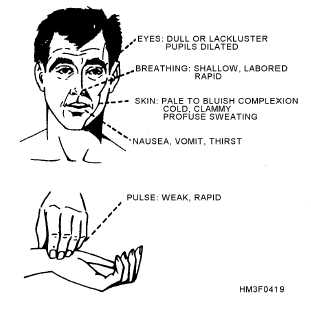
By constricting the blood vessels, we can maintain a reasonable blood pressure and adequate flow even with a smaller circulating volume. We normally vasoconstrict in the periphery — particularly the outer extremities and skin — “stealing” blood from those less-important tissues and retaining it in the vital core. This causes pallor (paleness) and coolness of the external skin. The sympathetic stimulation may also cause diaphoresis (sweating), which is not compensatory, but simply a side effect of the adrenergic release.

The heart also kicks into overdrive, trying to keep the remaining volume moving faster to make up for the loss. It beats faster (chronotropy) and harder (inotropy), resulting in tachycardia. Note that patients who use beta blockers (such as metoprolol) may not be able to muster much, if any, compensatory tachycardia.

A narrowing pulse pressure (the difference between the systolic and diastolic numbers) may be noted; since the diastolic reflects baseline pressure and the systolic reflects the added pressure created by the pumping of the heart, a narrow pulse pressure suggests that cardiac output is diminishing (due to loss of preload), and that more and more of the pressure we’re seeing is simply produced by shrinking the vasculature.

Tachypnea (rapid respirations) are also typically seen. In some cases, this may be due to emotional excitement, and there is also a longstanding belief that it reflects the body’s attempts to “blow off” carbon dioxide and reduce the acidosis created by anaerobic metabolism. (Interestingly, lactate — a byproduct of anaerobic metabolism — can be measured by lab tests, and is also a sign of shock, particularly useful in sepsis.) Additionally, it ensures that all remaining blood has the greatest possible oxygenation. However, it is also plausible that this tachypnea serves to assist the circulatory system: by creating negative pressure in the thorax (the “suction” you make in your chest whenever you inhale) and positive pressure in the abdomen (due to the diaphragm dropping down), you “milk” the vena cava upward during inspiration, improving venous return to the heart and allowing greater cardiac output. This “bellows” effect helps the heart fill more and expel more with each beat.

The more functional the patient’s body is — such as the young, strong, healthy victim — the more effective these compensatory systems will be. Hence the old truism that pediatric patients “fall off a cliff” — they may look great even up through quite profound levels of shock, due to their excellent ability to compensate, then when they finally run out of room they’re already so far in the hole that they become rapidly unhinged. It’s great that these people can compensate well, but it does mean we need to have a high index of suspicion, looking closely for signs of compensation (such as tachycardia) rather than outright signs of shock — because by the time the latter appears, it may be very late indeed.

Patients in compensated shock may become orthostatic; their bodies are capable of perfusing well in more horizontal postures, but when gravity pulls their remaining blood away from the core, this added challenge makes the hypovolemia noticeable. Less acute shock due to causes like dehydration may result in dry skin (particularly the mucus membranes; try examining the inside of the lower eyelid) with poor turgor (pinch a “tent” out of their skin and release it; does it snap back quickly or sluggishly?), and potentially with complaints of thirst. Urine output will usually be minimal. Generally, the more gradually the hypovolemia sets in, the more gradually it can be safely corrected; it’s the sudden, acute losses from causes like bleeding that we’re most worried about.


Decompensated Shock

As shock continues, compensatory systems will struggle harder and harder to maintain perfusion and pressure. Eventually they will fail; further vasoconstriction will reduce rather than improve organ perfusion, beating the heart faster will expel less rather than more blood, and the blood pressure will start to drop.

The hallmark of this stage of shock is the normal functioning of the body beginning to fail. The measured blood pressure will decrease and eventually become unobtainable. Pulses will weaken until they cannot be palpated. As perfusion to the brain decreases, the patient’s mental status will deteriorate. Heart rate and respirations, previously rapid, will begin to slow as the body loses the ability to drive them; like a government office that can’t pay its workers, the regulatory systems that should be fighting the problem begin to shutter their own operations. As the heart continues to “brady down,” eventually it may lose coherence (ventricular fibrillation), or keep stoically trying to contract until the last, but lose all effective output due to the lack of available blood (PEA). Cardiac arrest ensues, with dismal chances for resuscitation.


Alternative Forms of Shock

Although we have focused so far on hypovolemic shock, particularly of traumatic etiology, there are other possibilities. A wide range of shock types exist, but speaking broadly, there are only two other categories important to us: distributive, and cardiogenic/obstructive.

Distributive shocks include anaphylactic, septic, and neurogenic. The essential difference here is that rather than any loss of fluid, the vasculature has simply expanded. Rather than squeezing down on the blood volume to maintain an appropriate pressure, the veins and arteries have gone “slack,” and control of the circulating volume has been lost; it’s simply puddled, like standing water in a sewer pipe. (Depending on the type of shock there may also be some true fluid losses due to edema and third-spacing.) Imagine tying your shoes: in order to stay securely on your feet, the laces need to be pulled snugly (not too tight, not too loose). If the knot comes undone and the laces lose their tension, the shoe will likely slip right off. Your foot hasn’t gotten smaller, but the shoe needs to be hugging it properly to stay in place, and it’s no longer doing its job.

The hallmark of distributive shock is hyperemic (flush or highly perfused) rather than constricted peripheral circulation. The visible skin is warm (or hot) and pink (or red), and the patient may be profoundly orthostatic. Septic shock is associated with infection; anaphylactic with an allergic trigger; and neurogenic with an injury to the spinal cord.

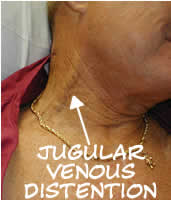
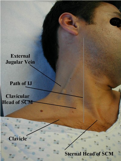
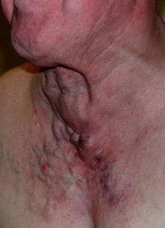
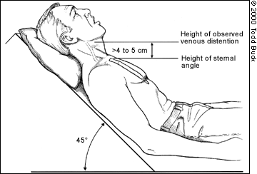
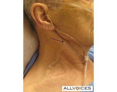
Cardiogenic and obstructive shocks are a different story. In this case, there’s nothing wrong with the circulating volume, or with the vasculature it flows within; instead, there’s a problem with the pump. Cardiogenic shock typically refers to situations like a post-MI heart that’s no longer pumping effectively. Obstructive shock refers to the special cases of pericardial tamponade, massive pulmonary embolism, or tension pneumothorax: physical forces are preventing the heart from expanding or blood from entering it, and hence (despite an otherwise functional myocardium) it’s unable to pump anything out. In either case, we can expect a clinical picture generally similar to hypovolemic shock, but likely with cardiac irregularities — such as ischemic changes or loss of QRS amplitude on the ECG, irregularity or slowing of the pulse, or changes in heart tone (such as muffling) upon auscultation. Pulsus paradoxus (a drop in blood pressure — usually detected by the strength of the palpable pulses — during the inspiratory phase of breathing), electrical alternans (alternating QRS amplitudes on the ECG), and jugular vein distention also may be present in the case of tamponade or severe tension pneumothorax.


In sum, remember these general points:

  1. The history and clinical context should be enough to make you suspect shock even without other signs or symptoms.
  2. The faster the onset, the more urgent the situation; acute shock needs acute care.
  3. Look both for signs of compensation (such as tachycardia) and for signs of decompensation (such as falling blood pressure). However, remember that due to confounding factors (such as particularly effective or ineffective compensatory ability, or pharmacological beta blockade), any or all of these may be absent.
  4. Distributive shocks are mainly characterized by well-perfused peripheral skin; cardiogenic/obstructive shocks are characterized by cardiac irregularities.

Interested parties can stay tuned for a brief appendix discussing fluid choices for resuscitation — otherwise, this journey through shock is finally finished!


Go to Part X (appendix) or back to Part VIII

The “Big Picture” Diagnosis

Our topic for today: diagnosis using a broad constellation of indicators, not a single red flag.

To mix things up, rather than read about it, let’s talk about it.

Here’s the quote I mentioned, from TOTWTYTR at the CCC blog.

Thoughts from WMEMS

This past weekend, I was able to attend the Western Massachusetts EMS Conference alongside such luminaries as Scott Kier and Kyle David Bates (of the extraordinary Pedi-U podcast). We sat through two days of outstanding lectures on various EMS-related topics, and walked away with some ideas and information I haven’t found anywhere else. Here are just a few of the unique pearls from the conference. Thanks to everyone for the great time!


Kyle David Bates on Mechanism of Injury

  • In an MVC, ejected (that is, fully ejected) victims have a 1/3 chance of a cervical spine fracture.
  • They also have around 25 times higher chance of mortality than an equivalent non-ejected patient.
  • Is “another death in the same vehicle” a legitimate concern when considering mechanism? Yes, but make sure that death wasn’t from an localized cause—for instance, a girder in the face, or they had a heart attack before they crashed.
  • How about “intrusion”? Over twelve inches into the patient compartment where your patient is found (meaning, visible from inside—not from the outside, which includes the buffer space of the walls), not including areas like the hood, trunk, etc. Alternately, over 18 inches into the patient compartment in areas where your patient is not found—for instance, the rear seating area, when you’re treating the solo driver.
  • “Distracting injuries” can mean painful injuries that distract the patient, but also gross stuff that distracts the provider. Consider a head-to-toe on virtually everyone, even when the funky arm fracture is drawing your attention.
  • Many “trauma” patients are no longer being treated with surgery anyway, so sending everything to the trauma centers overloads them for no reason.
  • One more reason why the sternal rub is not a great diagnostic: if they do clutch at their chest in response, is that localizing—or an abnormal, decorticate flexion response? Different GCS scores, but you can’t tell.
  • Are extremity injuries significant mechanisms? Penetrating injury proximal to the elbows or knees should be considered threatening to the torso, so yes. Pelvic fractures? For sure. (“How much blood can you lose into your pelvis? All of it!”)
  • With the automobile safety technology available today, you can crash fast, turn your car into a paperweight, but walk away unharmed. We no longer care about “high-speed,” only “high-risk,” which has many factors (see the Rogue Medic’s recent post on this).
  • Auto vs. pedestrians: kids get upper body injuries; adults get lateral trauma as we turn and try to get out of the way. Both can get run over.
  • Motorcycles. Harley-type riders seem to have more head injuries: they get hit by cars, due to low profile and dark clothing, and they wear partial helmets. Sports bikes get more extremity injuries: they wear good protection, are higher visibility, but they ride fast and run into things, breaking any and every bone they have.
  • Rollovers: no longer trauma criteria. You can roll and do great if you’re restrained. Number of rolls, final position, even roof intrusion have no correlation to injury severity.
  • Extrication time >20 minutes: no longer trauma criteria. Sometimes it just takes a while due to weather, access, etc, and newer vehicles are supposed to crumple more anyway.
  • Are burns trauma criteria? No. If they need specialized care, it’s a burn center, but this is not that time-sensitive—more a long-term management thing—so someone with burns and trauma should go to the trauma center instead, can be transferred later for burn care.
  • Helicopter transport: costs can range from $2,000 to $20,000 depending on distance, and insurers are refusing to pay many of these bills due to lack of necessity. Also consider the possibility of everyone dying in a fiery crash. Weigh cost vs. benefit.

Kyle David Bates on Shortness of Breath

  • Anxiety is caused by hypoxia; the cure for this is supplemental oxygen.
  • Sleepiness is caused by hypercapnia; the cure for this is bagging.
  • OPA or NPA? Testing the gag reflex may create a bigger airway problem (vomit). Better yet, check the mouth for pooled saliva; if present, there is no gag, use an OPA. If absent, they have a gag and are managing their own secretions, use an NPA.
  • Respiratory distress means there’s a problem, but they’re compensating (compensatory signs like tachypnea).
  • Respiratory failure means they’re decompensating (hypoxic/hypercarbic signs like altered mental status, cyanosis, falling sats)
  • Respiratory arrest means they’re not breathing.
  • Normal inspiration:expiration cycle about 1:2. Obstructive pulmonary problems impede expiration first, because that’s the passive process—it’s easier to inhale past obstructions because it’s an active process. So asthmatics have ratios like 1:4 or 1:5, they’re using active exhalation, and using auto-PEEP maneuvers. (Pursed lips in adults, grunting in kids.)
  • In adults, look for retractions intercostal (between the ribs) and sternal notch (between the clavicles); in kids, look substernal (below the ribs).
  • 40% of patients hospitalized with asthma have a pneumothorax! (Not necessarily clinically significant, though.)
  • Pulsus paradoxus/paradoxical pulses are a useful early sign of significant pulmonary dysfunction.
  • 90% of asthma attacks linked with an allergic reaction; however, rhinovirus (the common cold) may now be a contender. Others include: exercise (not sure why; maybe the temperature differential), active menstruation (asthma very common in young post-pubescent women—maybe the hormones), psychological (stress, panic), aspirin use.
  • Kids compensate great, so cyanosis (a decompensation sign) in kids is very late and very bad.
  • Risk-stratify these patients, because high risk patients can decompensate fast even if they look okay now. Previous hospitalizations? ICU admits? Intubations?
  • Cough asthma: no dyspnea, just dry coughing. It happens.
  • Smokers: measured in pack-years. 1 pack a day for 20 years is 20 pack-years, 2 packs a day for 5 years is 10 pack-years; 30–35 pack-years is where we start to see bad dysfunction.
  • Best place to check skin? Under the lower eyelid—lift it and check the mucus membranes. Dry for dehydration, pale for shock, blue for cyanosis, the whole gamut.
  • Ascites is a sign of fluid overload; try the fluid wave test. (Scroll down to “Examining for a fluid wave” here.)
  • Nebulized ipratropium/Atrovent: its role is mainly to reduce mucus and secretions (cf. atropine). Tachycardia etc. is not a contraindication, because it’s not absorbed systemically; it remains in the lungs.
  • Give nebs by hand-held mask or T-piece instead of strapping it to their face; that way you have a warning of deterioration when they can no longer hold it to their face.
  • Bronchodilators may not work great in beta-blocked patients.
  • Steroids take hours to have an effect, but the earlier they’re given the better the outcomes; give ’em if you have ’em.
  • If they need RSI, ketamine is nice because it also bronchodilates.
  • “Facilitated intubation” (i.e. snow ’em with a ton of benzos/narcs)? Be careful, because if you don’t get that tube, it’ll take forever to wear off; these aren’t short-duration drugs.

Kyle David Bates on Pediatrics

  • Use the Pediatric Assessment Triangle! Appearance, Work of Breathing, Circulation.
  • Appearance: General activity level and impression. Muscle tone, interactivity and engagement, look/gaze, crying. Appropriate appearance depends on age. Indicates a CNS/metabolic problem. (Make sure to check their sugar.)
  • Work of Breathing: Flaring, retractions, audible sounds, positioning. Remember they’re belly breathers.
  • Circulation: mostly skin. Cyanosis (bad), pallor, mottling (pallor + patchy cyanosis), marbling (in newborns—bright red skin with visible blood vessels, maybe some white areas—this is normal). Check cap refill on bottom of foot in little kids.
  • Shock in kids is most often from dehydration.
  • Airway: crying is a great sign. Remember to pad under the shoulders when lying flat, their huge heads can tip them forward and block the airway. Avoid NPAs in infants. In very small kids, breath sounds can transmit, so you may hear upper sounds in the chest or chest sounds in the trachea.
  • Under 2 months: peripheral cyanosis is normal, central cyanosis is bad. Limited behavior, often won’t visually track. Ask parents if their behavior is normal. Ask about obstetric history, it’s still relevant. They have no immune system really, so any infection (temp over 100.4) is a serious emergency.
  • 2–6 months: social smile, will track visually, recognize mom, strong cry and can roll/sit with support. May still be okay with strangers, but try to keep them with parents; if parents like you, they’ll like you
  • 6–12 months: stranger anxiety (unless they’re raised very communally). Very mobile and explore with their mouth, so always think about foreign body airway obstructions, especially up the nose, especially for dyspnea with sudden onset. Separation anxiety, so keep with parent. Offer distractions (toys, etc.). Do exam from toe to head so they get used to you before you reach their face.
  • 1–3 yrs (toddlers, “terrible 2s”): mobile, curious, opinionated, ego-centric, can’t abstractly connect cause-and-effect but learn from experience. Keep with the parents, distract them, assess painful part last (or everything you touch afterwards will hurt). May talk a lot or not much, it’s all normal, but they always understand more than they let on, so be careful what you say.
  • 3–5 yrs (preschool): magical thinkers, misconceptions (“silly” ideas like if they leak too much they’ll run out of blood), many fears (death/darkness/mutilation/aloneness), short attention span. Explain things in simple terms, relate to them (any cartoons or toys in the house you recognize?), use toys, involve them (here hold this, which arm should I use, etc). Don’t ever negotiate, just tell them what to do; praise them often; never ridicule.
  • 6–12 yrs (school aged): talkative, mobile, may not get cause and effect, want reassurance, involvement, praise. Live in present, may not think about danger or risk. Peer involvement. Speak directly to them, anticipate questions (will this hurt? am I going to die?), give simple explanations, don’t ever lie, respect privacy. If you need to do something painful (IVs, etc.) don’t tell them until just before, or they’ll dwell on it. Head-to-toe okay.
  • 13–18 (adolescents): regress when hurt or sick—act like big toddlers. Can understand and theoretically have common sense, but still take risks. Peer support. Speak directly, give concrete explanations, respect privacy, have patience.
  • Under 21 usually considered “pediatric.”
  • Degree of fever temp not associated with severity. No actual danger to brain until 106–107 degrees F or so.

Dr. Lisa Patterson on Trauma and Field Triage

  • RR <20 in infants is trauma center criteria since this is the one easily-measurable vital sign for them.
  • Crushed/degloved/mangled extremities: although not life-threatening, still worth the divert, because usually needs multi-specialty care (plastic surgery, orthopedics, hand specialists, etc.) to maximize function.
  • Calling in “altered mental status” or “unresponsive” is not super helpful—give a GCS or otherwise specify what you mean, there’s a big range here.
  • Trauma activations here are typically three tiers: category 1 (life threat), category 2 (no immediate emergency, but some concern or suspicion due to mechanism or presentation), consult (no concern on initial presentation, but later decision to admit, trauma paged down to consult).
  • Activation may alert/standby numerous parties including radiology, OR, pharm, blood bank, lab, ICU, respiratory, anesthesiology, social workers, etc. Not a small thing.

Sean Dorr on OEMS investigations

  • [This is Massachusetts-specific information; local providers can contact me directly if they want to hear about some of this material.— ed.]

Ginnie Teed on Organ and Tissue Donation

  • Donation is hugely hugely valuable and lifesaving, but there’s not nearly enough. About 60-70% of Americans are registered donors, around 100 million people, but only 1% end up as usable donors and we need far more. Low rates aren’t from consent, they’re from the logistics of getting viable candidates.
  • Uniform Anatomical Gift Act (UAGA) is federal regulation providing basic requirements for process; states use this standard to form their own systems. Registered donors must be recognized and organ procurement agencies are required to advocate for them even against wishes of family, etc. Driver’s license “opt-in” now considered legal consent in some but not all states.
  • National Organ Transplant Act establishes the rules of the registry, blinds the entire process, prevents manipulation or line-jumping; the database is centralized and controlled; you can’t legally buy or otherwise get around the system. Manipulation is taken very very seriously and massively investigated, because it’s not only unethical, the pall it casts over the process makes others decide not to donate—the result is many lives lost.
  • Referrals (i.e. calling procurement organization to say, “we have a potential donor”) come from hospitals, nursing homes, clinics, whomever. This process is exempt from HIPAA.
  • Tissues tested more heavily than organs, because if an infection is carried through transplanted (i.e. nonliving) tissue, it’s almost impossible to eradicate.
  • Organs used: vital organs. Heart, lungs, kidneys and livers (most common), pancreas, sometimes small bowel. Max 9 organs per donor.
  • Tissues used: not living, usually good for about 24 hours after death. Bones (not marrow, which is living), although we try to not obviously mutilate people (for their family’s sake), skin (hugely beneficial), corneas, vessels, heart valves, pericardium, connective tissue (for orthopedic repairs).
  • Three ways to declare death: neurological (no brain activity; body only alive due to our mechanical support; recovery team responds to site and performs planned recovery); cardiac death (heart stops; not planned); planned extubation/cardiac death (patient is mechanically supported, determination made that there is no possibility to survive on their own; vent is pulled, if heart stops within 59 minutes they can take some organs; usually just the durable liver and kidneys unless bypass is available).
  • Live organs can only be taken from perfused patients. Someone “dead” (i.e. no pulses) can be a tissue donor but not an organ donor unless you get ROSC. No point in continuing CPR to “maintain the organs” if there’s no possibility of getting return of circulation.
  • EMS documentation absolutely critical for determining donor eligibility. Need to know downtime in arrests, how much CPR, any ROSC no matter how brief, events/mechanism leading to arrest. There are hard limits on fluid/blood/colloids received, so they must know how much fluid you gave (reasonable estimate is fine). Must document all needlesticks, number and location; if they find any holes that aren’t accounted for they’ll have to assume they’re a drug user or that additional lines were started and extra liters given. If you don’t want to document something at least tell the receiving staff.
  • If blood is drawn, label must be placed so that expiration date of tube is still readable (FDA requirement).
  • Every donor can save up to 200 people; failure to document can kill just as many.

UMass Memorial LifeFlight on Air Ambulance Transport

  • Consider: how do you want the helicopter used? Need their higher level of care? Rapid transport to trauma center? Transport multiple patients in an MCI to more distant hospitals to reduce burden on closest facilities? Can even split the crew to provide higher level of care for multiple ground ambulances.
  • Many services simply will not fly into a hazmat situation.
  • Best makeshift landing zones are schools—big open areas, everyone knows where it is.
  • Wires are a major hazard, make sure to warn pilot—you can see them but he can’t.
  • Need about 100 x 100 ft for an LZ, or 35–40 big-ish strides per side. Secure the area against bystanders.
  • Hazards to clear, alert the pilot to, or just pick another spot: poles, antennas, trees, bushes, livestock, stumps, holes, rocks, logs, mile markers, debris. Tall grass can hide hazards. Close all vehicle doors, put your chinstraps on, secure loose items. Don’t stare at the bird landing, turn your back and watch for hazards.
  • Bad surfaces are dust, dirt, snow, ice, hay. Snow should ideally be very fluffy or very packed. If they land and get iced they may not be able to take off again. Don’t wash down a dusty LZ unless pilot requests it. Paved areas are simplest and best. Large clear roadways can land multiple choppers in a row.
  • Lighting options: orange traffic cone at each corner, with a handlight placed in each at nighttime. Or, flashing ministrobe at each corner. Or, vehicle headlights crossing the LZ. Don’t shine anything up at the helo, don’t mark with loose material, don’t use flares.
  • Designate one person as LZ Command (not the IC). Nobody else communicates with the helicopter. Your portable radio probably won’t reach them; use the mobile in the truck. If there’s any hazard on final approach, say one word—”STOP”—and pilot will abort.
  • Most crashes are pilot error, and most pilot error is due to fatigue. There should be hour limits for a pilot, and this is a valid reason to refuse to fly.

Detective John LeClair, EMT-P, on Opiates and Prescription Pills

  • Heroin is still big, but pills are a huge player now too. You get an easy prescription from a walk-in clinic or ED, pay maybe a couple bucks with Medicare/Medicaid, and can not only sell them for easy cash but can crush and snort/shoot it for the same effect as heroin. Then if money or access runs low, you end up on heroin anyway to chase that high.
  • Oxycontin/oxycodone best selling narcotic in the nation ten years ago, but now on the wane. You scrape off the time-release coating, crush it and snort or chew it. “Hillybilly heroin,” “blue,” “oxycotton,” “kicker,” etc. Street price about $1/mg (40mg, 80mg, 160mg common), so many turned to crime. In Aug 2010, manufacturer (Purdue) added a “geling” agent which turns it to gel when it contacts water, making it difficult to snort. Try to snort this Oxycontin OP and it turns into a ball in your nose. Some people are sticking straws/tubes up in there to try and get it deeper and deeper, so airway obstructions are happening.
  • Percocet: oxy plus acetaminophen. For years the most common analgesic for sports injuries, so common among youth. Kids shared ’em, put out bowls of them at parties, girls prostituted themselves for pills. Taken with alcohol the APAP/Tylenol kills your liver. “Littles,” “little babies,” “little dogs.”
  • Opana/oxymorphone: getting popular after Oxy OP started ruining everyone’s fun. Same idea but you can still snort it. Twice as strong, and costs twice as much ($2/mg)
  • How to grind? Take a hose clamp, cut it, straighten it, tape it down, run the pill across the holes to grind it. Or use a Pedi-Egg, which collects the powder for you. The finer, the better high.
  • Heroin: snort, “skin pop” (subcutaneous), mainline. Must be pretty pure to snort, which it now tends to be, so popularity grew (people were afraid of needles due to HIV). However now some HIV/Hep is spreading through bloody noses and sharing straws anyway.
  • Smack, horse, china white, chiva, junk, H, tar, black, fix, dope, brown, dog, food, negra, nod, white horse, stuff. Dealers have their own “brand names.”
  • Heroin addicts are creatures of habit; get high same place, same way. Any change in their routine (e.g. different location) can get them amped up, changing their sensitivity and leading to OD even with their usual dose. Consider this if you find an OD somewhere like a car or alley.
  • “Cotton fever”: they pluck out wads of cotton from cigarette filters and drop it in the heroin to help filter it. Sometimes when they draw out the liquid they get a bit of cotton, and when they shoot it they get a sort of phlebitis/infection/sepsis.

Differentiating Syncope: A Few Pearls

Syncope. To a fresh-faced student, it’s a snappy word for fainting. To someone with experience, it’s a heavy sigh, because we take a lot of calls for “syncope” and most of them are no big deal. But to a veteran provider, syncope is a deep, dark diagnostic hole—because syncope can be caused by countless different disorders, and although some are benign, a few of them are deadly.

Comprehensive diagnosis and treatment of syncope deserves its own dedicated series, and one of these days we’ll try and work through it from A to Z. Every etiology is unique and has its own distinct pathophysiology, presentation, and treatment considerations. Syncope sucks.

But for now, we’ll just talk about a few take-home pearls that can pay dividends in the everyday management of your next syncope call. We don’t support simplistic rules of thumb ’round these parts, but sometimes 95% of the work can be done by 5% of the know-how, and that’s just fine.

Here are a few dead-simple roadsigns to help guide you through the most common and most important causes of syncope.


Did they pass out and fall, or did they fall and then pass out?

Syncope means that somebody passed out and fell down. It doesn’t mean that they fell down and then lost consciousness. If they tripped on an oil can, fell over and smacked their head on a rock, they may have blacked out, but there’s no mystery there—it’s a simple trauma call.

So, our first step should be to take the raw he passed out and sift it into a more precise description. One problem is that people who lose consciousness often have a poor or unreliable memory of those events, so they may not always be helpful; this is why it’s nice to have witnesses who can tell the story. Of course, witnesses aren’t always reliable either.


Okay, so what do they remember?

To the extent that the patient remembers it, how do they describe the event?

A prodrome is an early, sometimes subtle set of symptoms that warn of a problem developing. Prodromes are our friend, because although they can be very brief or non-obvious, when present they can help indicate what happened. So, ask! It’s the O in OPQRST, and it’s the E in SAMPLE, so it’s the beginning and end of our patient history—no excuses!

Vasovagal syncope is one of the most common causes of syncope, involving a transient drop in blood pressure, and vasovagal syncope is usually preceded by a prodrome. If you’ve never had the experience of standing up too fast and getting briefly faint, here’s the gist: you become light-headed, your vision blurs or darkens, you feel weak, you may stumble, and finally you go down. There may also be broad neurological symptoms, such as visual disturbances (“seeing spots”), strange sensations, shaking, and more. (Basically, your brain isn’t getting enough oxygen, so odd stuff happens.)

How about seizures? Many seizures are preceded by a prodrome known as an “aura,” which can manifest as various unusual neurological abnormalities; read more in our piece on seizures. Did the patient truly lose consciousness, or do they claim that they remained somewhat aware? In a simple partial seizure, the patient will remain aware of their surroundings (although these often don’t cause a “syncopal” collapse); in most others they will experience a gap in consciousness.

Syncope caused by cardiac arrhythmias, such as a run of V-tach or a Stokes-Adams attack, will sometimes be preceded by a palpable sensation of weakness, or palpitations  (“fluttering”) in the chest. However, in many cases there will be no warning whatsoever.


What did the witnesses see?

It’s one thing to hear about a prodrome from the patient, but you may get a different story from the bystanders.

What did they see before he went down? Did he become absent, demonstrate tics or tonic immobility, perhaps complain of an aura? Did he demonstrate obvious clonic jerking of the muscles or urinary incontinence? If he’s acting normally now, was there a period after the event where he demonstrated sluggish activity or unusual behavior, consistent with a post-ictal period? These are all suggestive of a seizure.

Were his eyes open or closed for the duration? Closed is typical of classic syncope, such as a vagal event; open is more appropriate for a seizure. If open, were they rolled back? This also suggests seizure.

Did the patient say, do, or complain of anything before or after the event, which he may no longer recall? Dizziness, headache, chest pain?

Did he stumble, lean against something, or seem to become dizzy? After he went down, did he regain consciousness almost immediately? These are suggestive of vasovagal; once a horizontal position is reached, perfusion to the brain is restored and the problem resolves. If he remained unconscious for a prolonged period while prone—or his initial episode occurred while already seated or reclined—this is highly unusual for vasovagal.

Was he walking and moving normally, in no distress, when he suddenly collapsed like a marionette with its strings cut, hitting the ground with no attempt to protect himself? This is strongly suggestive of a cardiac event and these patients should be considered high-risk for sudden death.


Is there a suggestive history or surrounding circumstances?

Sometimes, the chain of events or the patient’s medical history may suggest an etiology.

Is there a known history of a seizure disorder like epilepsy? How about diabetes? (Take a blood sugar if you’re capable of it; in my book, everybody with an altered mental status is diabetic.) Do they have often pass out or become light-headed?

Have they been eating and drinking as normal? Have they had the flu, and been unable to keep down fluids for the past two days? Were they partying all night? Vomiting? Are they a marathon runner who collapsed in 110 degree weather? Dehydration is a common cause of syncope, particularly in the young, healthy population.

Is there a known condition which may have neurological or metabolic involvement? Cancer with metastases to the brain? A recent infection? A congenital heart condition, such as Long QT, hypertrophic cardiomyopathy, or Brugada? For that matter, are they currently drunk or using drugs? If they take psychotropic or other medications, are they compliant with these, or could there have been an under- or over-dose?

Has there been any recent trauma, such as a fall, motor vehicle collision, or assault with injury?

Have there been repeated lapses in and out of consciousness, rather than a single event? This is an ominous sign suggesting a significant problem.


Are there frank clinical signs that suggest a diagnosis?

This is less likely to be useful than the history, but it can help rule in or rule out major, acute emergencies.

Cardiac abnormalities may manifest with irregular pulses, and active decompensation may be revealed in the blood pressure. Whenever possible these patients should receive ECG monitoring, including a 12-lead. Orthostatic vital signs can be considered if vagal, orthostatic, or hypovolemic etiologies are suggested.

All syncope patients, including suspected seizures, should get a neurological workup, particularly a Cincinatti Stroke Scale.

Respiratory adequacy, including pulse oximetry where available, should be assessed.

Evaluate the abdomen for signs of hemorrhage, and inquire about blood in the stool or emesis as well.

What it Looks Like: Jugular Vein Distention

See also what Agonal RespirationsSeizures, and Cardiac Arrest and CPR look like

Jugular vein distention or JVD (alternately JVP — jugular vein pressure or jugular vein pulsation) is right up there among the most mentioned but least described clinical phenomena in EMS. If you tried to count how many times it occurs in your textbook, you’d run out of fingers, but many of us graduate without ever seeing so much as a picture of it, never mind developing the acumen to reliably recognize it in an emergency.

JVD is simply the visible “bulging” of the external jugular veins on either side of the neck. These are large veins that drain blood from the head and return it directly to the heart. Since they’re located near the surface, they provide a reasonably good measure of systemic venous pressure.

JVD is elevated any time venous return is greater than the heart’s ability to pump the blood back out. Remember that we’re not talking about the vessels that plug into the left heart; that involves the pulmonary arteries and veins, which are not visible in the neck. (Instead, the best indicator of pulmonary hypertension is audible fluid in the lungs.) Rather, we’re talking about the systemic vasculature, which drains into the right ventricle via the right atrium. When veins aren’t getting emptied, we look downstream to discover what portion of the pump is failing. JVD is therefore caused by right heart failure. (Of course, the most common cause of right heart failure is left heart failure, so that doesn’t mean it’s an isolated event.) If JVD isn’t the heart’s fault, then we look to fluid levels. Too much circulating volume will lead to bulging veins for obvious reasons; the flexible tubes are simply extra full.

Although it’s probably most often seen, and most diagnostic, in volume-overloaded CHF patients, the main reason JVD is harped upon in EMS is because it’s a useful sign of several acute emergencies. Mainly, these are obstructive cardiac conditions, where some sort of pressure is impeding the heart’s ability to expand, and immediate care to relieve the pressure is needed in order to prevent incurable deadness. Much like the bladder, the heart is just a supple bag of squishy muscle, and although muscle is very good at squeezing, it has no ability to actively expand. The heart therefore fills only with whatever blood passively flows into it, and if it’s being externally squeezed by pressure in the chest, it can’t fill very much.

Tension pneumothorax is perhaps the most common cause, where air leaks from the lungs into the chest cavity with no way to escape; as the pressure in the chest increases, it bears down on the heart. Associated symptoms are respiratory difficulty, decreased breath sounds on the affected side, and hypotension. Pneumothorax can be readily corrected by paramedics using needle decompression.

Cardiac tamponade is another cause, where fluid leaks from the heart into the pericardium, an inflexible sac that surrounds it (this leakage is called a pericardial effusion), eventually filling the available space and compressing the myocardium. Associated symptoms are hypotension and muffled heart sounds (these plus JVD are known as Beck’s triad). Tamponade cannot be treated in the field, but an emergency department can perform a pericardiocentesis, where a needle is inserted through the pericardium. (For the medics out there, electrical alternans on the monitor is also supportive of tamponade.)

A rather less common syndrome that can produce similar obstructive effects is severe constrictive pericarditis, inflammation of the pericardium usually caused by infection.

JVD is not an all-or-nothing finding — the amount of distention visible at the neck will depend on the degree of venous pressure. Gravity wants to pull blood back down, so the more venous pressure, the higher on the neck distention will climb; profound JVD reaches many inches up the neck, slight JVD will only cover a few centimeters. The pressure can actually be quantified by measuring the vertical height of the highest point of distention (measured from the heart itself, using the angle of Louis as a landmark), but this is probably more detail than is needed in the field. Suffice to say that distention reaching more than 2-4cm of vertical distance (as opposed to the distance on the neck) above the chest is usually considered pathological, and less than 1-2cm can be considered suggestive of hypovolemia.

If it changes with respiration, JVD should rise during expiration and fall with inspiration. Breathing in involves using your diaphragm to create “suction” in the chest, reducing pressure and allowing greater venous return — draining the jugulars. A paradoxical rise in JVD during inspiration (think: up when the chest goes up) is known as Kussmaul’s sign (not to be confused with Kussmaul respirations, which is a pattern of breathing), and is particularly suggestive of obstructive pathologies.

JVD can be difficult to appreciate in all but the most significant cases. It helps to turn the patient’s head away and illuminate the area with angled backlighting, which creates a “shadow” effect. Jugular pulsation should not be confused with a visibly bounding carotid pulse. To distinguish them, remember that although jugular veins may visibly pulsate, their rhythm is generally complex, with multiple pulsations for each single heartbeat (you can feel the carotid to compare the two). The jugular “pulse” will also never be palpable; the distention can be easily occluded by the fingers and will feel like nothing.

Strictly speaking, the internal jugular is usually considered more diagnostically useful than the external jugular, but it’s far harder to examine, so the latter is often used. For various reasons, many people also find the right jugular more useful than the left, although in an ambulance it’s harder to examine.

Most often, JVD is examined in an inclined or semi-Fowler’s position of 30-45 degrees. If the patient is supine, a total lack of visible JVD is actually pathological and indicative of low volume; in this position the jugular veins are usually well-filled. (Think: flat veins in a flat patient is bad.) JVD when the head is elevated is more to our interest.

Some examples of visible JVD follow, plus some examination tips. It is recommended that you start checking this on your healthy patients now, so you’ll know what it looks like before you try to make a diagnostic call using its presence. And until you do, stop documenting “no JVD” on your assessments!

Significant JVD

Significant JVD

A different, much larger view of the same (click to enlarge)

A different, much larger view of the same (click to enlarge)

Click through for a good discussion of JVD assessment

Click through for a good discussion of JVD assessment

Some more subtle JVD

Some more subtle JVD

The basic method of measuring JVD

The basic method of measuring JVD

A nicely thick and squiggly external jugular

A nicely thick and squiggly external jugular

Here’s a student making her external jugular “pop” by heavily bearing down, aka the Valsalva maneuver. This markedly increases thoracic pressure, increasing venous backup; it’s an exaggeration of the effect seen during normal exhalation.

Another example of someone inducing JVD by a Valsalva

Here’s a great video demonstrating the appearance of JVD, how to measure it, and testing the abdominojugular reflex (formerly known as the hepatojugular), which involves pressing down on the abdomen to raise thoracic pressure.

A brief clip of jugular venous pulsation, visible mainly toward the suprasternal notch.

http://www.youtube.com/watch?v=sOpn6_r7Wo4