Podcast: EMS to ED Interface

Streamlining a patient’s entry to the healthcare continuum is one of our main roles in EMS, and the key step in most cases is when we transfer care at the emergency department. This isn’t rocket science, but you can do it well or less well, and frankly I think it’s tough to do right unless you can see the whole picture. We never really know in what ways we’re setting up people effectively for their ED care and in what ways we’re part of the problem, unless perhaps we work on both sides.

So I asked for a little help here. I sat down virtually with Dr. Brooks Walsh, ED attending extraordinaire — author of Mill Hill Ave Command and Doc Cottle’s Desk — and with Jeff, an ED nurse from my area. We discussed how to work and play together better, including topics like handoff reports, useful histories, and typical ED courses of care.

Click here to listen or download (1:15, MP3 format)

A few of the bullet-worthy points:

  • Jeff’s hospital saves time in all trauma, stroke, and STEMI activations by assigning patients an alias immediately upon notification by EMS. That way registration isn’t lurking around while the team is trying to treat the patient.
  • Cath lab activations from the field are still often about trust — whether staff knows the individual provider or the particular service calling. Rightly or wrongly, there’s also a stricter de facto standard for activation during off hours when nobody wants to get out of bed.
  • For stroke, neurology may be in the room when you arrive, but more often, especially in smaller hospitals, they’re available by page or teleconference.
  • When bringing in the stroke, try and ensure that family who can testify to time-of-onset/time-last-seen-normal, as well as consent to treatment on the patient’s behalf, are present — ideally transported with you — not unavailable in a taxi somewhere.
  • When you walk in the room, the typical team is a doctor, a nurse, a tech, then any extras — residents or other students, surgery, pediatrics, whomever. And registration is the dude with the clipboard or computer, of course.
  • When reporting to the doc, focus on: first, anything that needs to happen immediately; second, information he can’t get elsewhere (i.e. not patient medical history unless it’s not available in the records, laundry list of negatives, etc.), such as how you found the patient, general context, changes en route, etc.
  • Written PCRs are usually not read due to difficulty obtaining them and general unfriendliness (hard to find info, obscure writing), but sometimes there’s useful stuff in there, particularly in the narrative itself.
  • Baseline patient info from EMS is great if we know the patient well (frequent fliers); baseline info from bystanders, staff, family etc. is okay but less reliable.
  • Get patients to their usual facility if at all possible, especially those with complex histories, and especially anyone with recent surgical history — otherwise they’ll just get transferred later.
  • “Take me to x, my doctor is there” (meaning PCP or specialist) — less important, but can be nice if there are chronic issues and they’d like to maintain the existing treatment plan.
  • Disagreements over patient triage or treatment: find the attending or perhaps resource nurse and voice your concern. In the long-term: raise issues with the hospital’s EMS liaison (either directly or through your internal chain of command).
Glucometry: Clinical Interpretation

Continued from Glucometry: Introduction and Glucometry: How to Do it

Implementing glucometry into your overall assessment means understanding three things: when to use it, what the results mean, and when it fails.


Indications

First of all, by and large the only people with derangements of their blood sugar should be diabetics. The rest of us are generally able to maintain euglycemia through our homeostatic mechanisms, except perhaps in critical illness causing organ failure and similar abnormal states. Now, if someone injected you — a non-diabetic — with a syringe of insulin, you’d become terribly hypoglycemic, since it would overwhelm your body’s ability to compensate for the loss of glucose. But nobody’s likely to do that if you’re not a diabetic, unless it’s meant for somebody else and a drug error occurs, or I suppose if they’re trying to assassinate you.

With that said, people walk around who are diabetic and don’t know it. I’ve lost track of the patients I’ve transported who presented with signs suggestive of a diabetic emergency, denied a history of diabetes, and came back with a BGL of 600. Well, my friend, I have some bad news for you. “Everybody is diabetic, even if they’re not” is my attitude. Almost a fifth of older Americans are diagnosed, and the older and sicker they are, the more common it is.

Which brings us back to: who needs a BGL?

The most correct answer is anybody with clinical indications of either hypo- or hyperglycemia. As we saw, diabetes itself is really associated with hyperglycemia, which is why the classic signs of hyperglycemia are usually used to diagnose diabetes: polyuria (excessive urination, as extra glucose is excreted by the kidneys and brings water along with it osmotically), polydipsia (excessive thirst and water consumption, to replace the fluids urinated out), and polyphagia (constant hunger, since despite all the sugar floating around it’s not reaching the cells very easily). If your patient is complaining of those, you might be the first one to discover their condition. The diagnosis doesn’t require elaborate tests and imaging; a fasting glucose over 126 BGL tested on multiple occasions, or just once in combination with clinical symptoms, or a post-prandial (after eating) glucose exceeding 200, is the definition of type II DM. (With that said, I wouldn’t go around diagnosing your patients; that’s not your job, and you’re not quite that good.)

Once the glucose gets higher than the “renal threshold” — usually around 180 in average folks — the body starts to excrete it into the urine. This can actually be detectable by chemical dip-stick, or even by odor and texture at very high levels.

When hyperglycemia becomes severe and prolonged enough, we start to worry about diabetic ketoacidosis. Although burning fat and protein is not necessarily dangerous (some popular diets actually put you into a mild ketogenic state intentionally), extensive accumulation of ketones caused by a total lack of insulin (as in type I diabetics — DKA is rarely seen in type II) creates a metabolic acidosis in the body. This is when the long-term harm of hyperglycemia becomes a short-term hazard. DKA causes altered mental status, usually elevated states of confusion and disorientation, and combative behavior isn’t uncommon. Combined with the acetone odor that sometimes presents on the patient’s breath — which can smell like alcohol — DKA patients can seem suspiciously like drunks, and treating them like drunks is a great way to go down a bad path. (A word of wisdom: not only is everybody diabetic, but drunks are definitely diabetic.) DKA also frequently presents with symptoms of dehydration, due to the osmotic water loss in the urine; nausea and vomiting; and deep, rapid Kussmaul breathing to blow off the acidic CO2.

A few situations can cause short-term hyperglycemia, including stressors of any kind (there’s even “white coat hyperglycemia,” where patients tend to produce elevated sugars at the doctor’s office), but these typically won’t produce anything like the massive levels leading to DKA.

With all of that said, you need to really build up some glucose before hyperglycemia becomes symptomatic, and even more than that before it becomes acutely dangerous and unstable. That’s why as a rule, we’re more concerned with hypoglycemia, usually due to medication administration, physical exertion, or metabolic demand exceeding what was expected. Hypoglycemia again presents as altered mental status, in this case more often an inhibited rather than an elevated state: confusion, lethargy, disorientation, inability to focus or follow commands, weakness, headache, seizures, and eventually coma and death. The fun part is that the impairments can present as focal as well as generalized deficits: unilateral weakness of the limbs or face, speech slurring, poor gait, vision abnormalities, and more. In fact, hypoglycemia is a neurological chameleon, and can look like almost anything; it’s particularly notorious for imitating strokes, and for causing (not imitating) seizures. Interestingly, kids are particularly prone to hypoglycemia due to their gigantic heads, full of glucose-hungry brain.

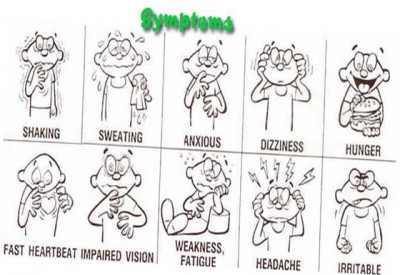
Despite all this, the primary manifestations of early hypoglycemia are actually not symptoms of hypoglycemia. Rather, they’re caused by catecholamines — by the body releasing stress hormones, primarily epinephrine, in a response to the emergency. (This is not an irrational move: epinephrine helps us release and retain glucose.) As a result, we often seen the same signs we’d expect in anybody with a profound sympathetic stimulus: pale and diaphoretic skin, anxiety and shakiness, tachycardia and hypertension, even dilated pupils. Wise diabetics recognize the early signs of this sympathetic response and drink some Pepsi. As levels keep dropping, these symptoms combine with the neurological effects of glucose starvation to produce a confused, sweaty, increasingly stuporous individual. If left untreated, finally the sugar drops until we’re looking at the picture of impaired and diminished consciousness caused by true hypoglycemia. So just like always, the signs of compensation are our early warning system; once the body decompensates, it’s already late in the game.

To make a long story short, anybody with altered mental status, or any kind of general systemic complaint (weakness, fatigue, anxiety, nausea, etc.) should probably get their glucose tested, whether or not they have a known history of diabetes. This is true even if you suspect another cause, such as stroke. Not only can diabetic emergencies look like anything, they can also be comorbid; it is extremely common for patients to have another problem, yet also to bring a high or low sugar along for the ride, due to the illness throwing a wrench in their normal intrinsic and extrinsic glycemic homeostatic systems.

A number of years ago, there was some limited but compelling research that suggested poorly-controlled blood glucose (meaning not severe derangements but merely small deviations from the ideal range) was associated with increased mortality among an inpatient population with a wide variety of conditions. In other words, if you were hospitalized with something like sepsis, you were more likely to end up dying if your sugar tended to float around 160 instead of 110. As a result, it become trendy to practice extremely tight and aggressive glucose management for virtually everybody; diabetic patients were being tested every few hours and ping-ponged around using medication to keep their numbers textbook-perfect. More recently a number of studies have suggested that this may be less important than was thought, and in fact that excessive paranoia leads to a lot of iatrogenic harm from accidental insulin overdoses. This battle is still being fought in the hospitals, but for our purposes a reasonable take-away would be: when managing acute illness, from sepsis to head injury to cardiac arrest, once everything else is done it’s not a bad idea to check the patient’s sugar.


What’s the Number Mean?

So you’ve taken a blood glucose, either by capillary finger-stick or from a venous sample. Now what?

We mentioned that the “normal” range is something like 70–140. Diabetics seeking to control their condition and not have their toes falling off in a few years usually strive for tighter control of their BGL than is needed for acute care; a sugar of 175 is a little on the high side for a routine check, but a pretty meaningless elevation for our purposes.

All things are also relative, in that a given BGL must be compared to the patient’s baseline to predict its effects. In other words, poorly-controlled diabetics who are routinely sitting at 200 may become symptomatic of hypoglycemia at relatively high levels, whereas very well-controlled diabetics who usually run lower may be able to drop very low indeed without noticing it. However, a few rules-of-thumb are useful:

Non-diabetics usually become noticeably symptomatic below a sugar of, on average, about 53. (Diabetics, particularly those who are usually poorly-controlled, are more variable — their average symptomatic threshold is more like 78.)

After a recent meal, diabetics may demonstrate hyperglycemia to various degrees depending on whether they ate a Cobb salad or an entire chocolate cake. Non-diabetics should not exceed 200 or so. A few people can exhibit hypoglycemia after meals, due to alcohol consumption, “dumping syndrome,” or some other phenomena, but far more often they’ll exhibit similar symptoms without any true hypoglycemia; some people get shaky and sick due to postprandial epinephrine release.

After an unusual period of fasting (“haven’t eaten since yesterday”), non-diabetics should still have a largely unremarkable sugar. For diabetics, it will depend mainly on how much and what type of medication they’re using.

There’s usually a gap of 10–20 mg/dL between hypoglycemia that’s noticeable to the patient (i.e. sympathetic effects) and hypoglycemia that causes cognitive impairment (i.e. neurological changes). This is their safety margin, when they’re taught to eat or drink some fast carbs; if it keeps dropping they may no longer be able to take care of themselves.

But here’s the problem: the sympathetic “warning signs” can be mediated or impaired for various reasons. For one thing, if your body has to flip that switch often, you become numbed to it, and your hypoglycemic thresholds becomes lower and lower. And many patients with various metabolic and endocrine failures simply can’t muster much of a stress response — the same reason why the elderly may not produce tachycardia and other shock signs when they become hypovolemic. Finally, drugs like beta blockers that directly block sympathetic activity can seriously obscure hypoglycemia. Grab your nearest bottle of beta blockers and read the list of adverse effects: one will be hypoglycemic unawareness, a five-dollar term that means beta blockade can make it difficult to know when your sugar drops low.

Another important consideration in evaluating glucose levels is the expected trend. For instance, a BGL of 70 in a diabetic patient might not excite anybody. However, if you’re testing her because her nurse said that she just accidentally received four times her normal insulin dose, then a BGL of 70 should be alarming, because it’s probably going to keep dropping, and she doesn’t have very far to go.

To make a long story short, the clinical effects of both hypo- and hyperglycemia can vary substantially. What to do? It’s simple: assess the patient physically, obtain a history of their oral intake, medications, and metabolic demands (such as exercise), test their sugar if there’s any possibility of glucose derangement, and compare all those data against each other. A low number in the setting of obvious clinical symptoms is bad. A low number in an asymptomatic patient, or a normal number in a patient with highly suggestive signs and symptoms, should force you to bring out your thinking cap and weigh the odds.

What about treatment? Severe hypoglycemia needs ALS or the hospital — they’ll receive IV dextrose. Severe hyperglycemia needs the hospital only, where they’ll receive carefully-dosed insulin; this is generally considered too dangerous to administer in the field (although patients may have their own), so paramedics are reduced to giving fluid boluses, which may help dilute high glucose concentrations (not a very elegant solution) and is probably needed by a patient in DKA anyway, but isn’t really a fix.

What about oral glucose, in the cute little tubes we carry? Typically these are gels containing 15g of glucose, taken orally (either swallowed or held in the mouth — against the cheek or under the tongue — until it’s absorbed). Do they work? Sure. But it’s not much sugar and it’s not very fast. I found one source that suggests 15g of oral glucose should raise the BGL by 50 mg/dL within 15 minutes of administration — but I’ve never found it to be nearly that effective. In my experience, a bump of about 10 mg/dL per tube is about the best you can hope for in the short-term. If you need more than that, go with the medics and the IV syrup.


Testing Errors

When is a tested capillary or venous glucose unreliable? Usually it’s your fault.

Well over 90% of BGLs that test outside the maximum error range (remember, around 15%) are due to user error. Some of the main ones:

  • Your meter requires lot coding, and you failed to do so or used strips from the wrong lot.
  • You failed to clean the skin before lancing, contaminating the sample (not to mention creating an infection risk), or you had some D50 on your glove and it got mixed in there.
  • Rather than gently wicking the sample into the strip, you “smeared” the two together with mechanical pressure, interfering with the expected reaction process.
  • You drew blood from an arm with an IV infusion of D50, TPN, or other meds distal to it. Particularly when peripheral perfusion is poor, always try to sample at a different limb from any running drips.
  • You tried to reuse a non-reusable strip (gross).

Okay, okay, so nobody’s perfect. Factors that may not be as obvious include:

  • Temperature. The test reaction is designed to function within a specific temperature range, which is broad (often around 40–104 degrees) but not limitless, so don’t use them in freezing weather, and try not to leave your equipment ungaraged without climate control when it’s very hot or cold out.
  • Altitude. Just in case you’re an Everest expedition doctor.
  • Humidity. The strips have trouble when it gets very humid.
  • Air. The reagents in the strips will actually degrade if exposed to air for sufficient periods of time, so make sure that you keep them in their tightly-sealed case, and follow their printed expiration dates.
  • Time. If you draw whole blood and leave it around (much more likely to happen in the laboratory than in the ambulance), the erythrocytes will metabolize glucose at about 5-7% per hour.

The good news is that in many of these situations, internal error-checking within the glucometer will recognize the problem, and flash an error rather than a reading. Errors messages are usually numbered and can be informative, but each manufacturer uses different codes, so read the manual if you want to know what “ER2” means. (Hint: not enough blood in the sample is by far the most common.) Many of the other problems can be caught if you regularly check the meter using a known-value test solution, which you should be doing anyway according to most drug and safety agreements. (By the way, both the test strips and those vials of solution are usually meant to expire a few months after opening — the printed date is for an unopened bottle — so if they’ve around forever it’s probably time to retire them.)

What about physiological states that can interfere with the reading? We’ve discussed a few, but briefly:

  • Hematocrit. Anemia from any cause, including cancer or blood loss, causes falsely high readings. High crit, common in neonates, causes falsely low readings.
  • PaO2. Oxygen interferes with the electrochemical redox reaction; thus high concentrations of dissolved oxygen cause falsely low readings, and low PaO2 (i.e. hypoxia) cause falsely high readings, potentially masking a true hypoglycemia.
  • pH. Primarily in meters using the glucose oxidase enzyme, alkalosis will cause falsely elevated readings, while acidosis causes falsely low readings. The acidosis of DKA can therefore cause falsely low readings, masking the profound underlying hyperglycemia, so if the clinical picture screams DKA, don’t necessarily let the glucometer tell you different.
  • Macronutrients. High levels of circulating proteins or fats can cause falsely low readings due to dilution.
  • Hypoperfusion and inadequate circulation. See our previous remarks on this, and remember that venous sources will be more accurate than capillary.

Finally, are there medications that can interfere with glucometer accuracy? There sure are. These in particularly are highly device-dependent, with the glucose oxidase-type meters most often affected. Generally, the effects are not profound, but occasionally they may be clinically relevant.

  • Ascorbic acid. Better known as Vitamin C, some people take megadoses of this stuff, thinking it’ll cure their cold or flu. Depending on the meter it can cause falsely high or low readings, usually a minimal change, but at “megadose” levels the effect can be significant.
  • Acetaminophen. Also known as Tylenol. The effect is similar to ascorbic acid, but even more modest; it should only be considered in major overdoses, and even then the difference is unlikely to break 35.
  • Dopamine. Massive doses, such as might be used for intensive inotropic support, can modestly influence glucose dehydrogenase-based meters.
  • Mannitol. High doses can elevated the measured BGL by around 35.
  • Icodextrin. This is a dialysate solution used for peritoneal dialysis (not hemodialysis — this is where they pump fluid into the abdomen, let it sit, then drain it out), mainly in patients with diabetes. It metabolizes to maltose, which can cause falsely elevated readings in certain meters. There’s at least one tragic and unfortunate case report of a patient death resulting from massive insulin overdose due to this effect, not noticed until the true BGL was obtained by laboratory analysis. If your patient undergoes peritoneal dialysis, try to find out what dialysate is used, and if that’s not possible, it may be safest to assume their sugar is lower than you’re measuring.


Conclusions

After all this you’re probably thinking glucometry is so convoluted and rife with pitfalls that you’re better off just eyeballing how sweet your patients are. But don’t let me turn you off! This remains one of the best assessment aids we have, because diabetic emergencies remain some of the most common, most treatable, and most easily confused disorders that we encounter. We can’t perform exploratory surgery, and we may never see prehospital CT scans, but this is a diagnostic test that’s so cheap and simple, with such real potential to affect your decisions, that it should be available everywhere. If you maintain your equipment, learn how to do it right, and keep a few basic confounders in mind, it’ll serve you well as one of your most reliable tools.

Differentiating Syncope: A Few Pearls

Syncope. To a fresh-faced student, it’s a snappy word for fainting. To someone with experience, it’s a heavy sigh, because we take a lot of calls for “syncope” and most of them are no big deal. But to a veteran provider, syncope is a deep, dark diagnostic hole—because syncope can be caused by countless different disorders, and although some are benign, a few of them are deadly.

Comprehensive diagnosis and treatment of syncope deserves its own dedicated series, and one of these days we’ll try and work through it from A to Z. Every etiology is unique and has its own distinct pathophysiology, presentation, and treatment considerations. Syncope sucks.

But for now, we’ll just talk about a few take-home pearls that can pay dividends in the everyday management of your next syncope call. We don’t support simplistic rules of thumb ’round these parts, but sometimes 95% of the work can be done by 5% of the know-how, and that’s just fine.

Here are a few dead-simple roadsigns to help guide you through the most common and most important causes of syncope.


Did they pass out and fall, or did they fall and then pass out?

Syncope means that somebody passed out and fell down. It doesn’t mean that they fell down and then lost consciousness. If they tripped on an oil can, fell over and smacked their head on a rock, they may have blacked out, but there’s no mystery there—it’s a simple trauma call.

So, our first step should be to take the raw he passed out and sift it into a more precise description. One problem is that people who lose consciousness often have a poor or unreliable memory of those events, so they may not always be helpful; this is why it’s nice to have witnesses who can tell the story. Of course, witnesses aren’t always reliable either.


Okay, so what do they remember?

To the extent that the patient remembers it, how do they describe the event?

A prodrome is an early, sometimes subtle set of symptoms that warn of a problem developing. Prodromes are our friend, because although they can be very brief or non-obvious, when present they can help indicate what happened. So, ask! It’s the O in OPQRST, and it’s the E in SAMPLE, so it’s the beginning and end of our patient history—no excuses!

Vasovagal syncope is one of the most common causes of syncope, involving a transient drop in blood pressure, and vasovagal syncope is usually preceded by a prodrome. If you’ve never had the experience of standing up too fast and getting briefly faint, here’s the gist: you become light-headed, your vision blurs or darkens, you feel weak, you may stumble, and finally you go down. There may also be broad neurological symptoms, such as visual disturbances (“seeing spots”), strange sensations, shaking, and more. (Basically, your brain isn’t getting enough oxygen, so odd stuff happens.)

How about seizures? Many seizures are preceded by a prodrome known as an “aura,” which can manifest as various unusual neurological abnormalities; read more in our piece on seizures. Did the patient truly lose consciousness, or do they claim that they remained somewhat aware? In a simple partial seizure, the patient will remain aware of their surroundings (although these often don’t cause a “syncopal” collapse); in most others they will experience a gap in consciousness.

Syncope caused by cardiac arrhythmias, such as a run of V-tach or a Stokes-Adams attack, will sometimes be preceded by a palpable sensation of weakness, or palpitations  (“fluttering”) in the chest. However, in many cases there will be no warning whatsoever.


What did the witnesses see?

It’s one thing to hear about a prodrome from the patient, but you may get a different story from the bystanders.

What did they see before he went down? Did he become absent, demonstrate tics or tonic immobility, perhaps complain of an aura? Did he demonstrate obvious clonic jerking of the muscles or urinary incontinence? If he’s acting normally now, was there a period after the event where he demonstrated sluggish activity or unusual behavior, consistent with a post-ictal period? These are all suggestive of a seizure.

Were his eyes open or closed for the duration? Closed is typical of classic syncope, such as a vagal event; open is more appropriate for a seizure. If open, were they rolled back? This also suggests seizure.

Did the patient say, do, or complain of anything before or after the event, which he may no longer recall? Dizziness, headache, chest pain?

Did he stumble, lean against something, or seem to become dizzy? After he went down, did he regain consciousness almost immediately? These are suggestive of vasovagal; once a horizontal position is reached, perfusion to the brain is restored and the problem resolves. If he remained unconscious for a prolonged period while prone—or his initial episode occurred while already seated or reclined—this is highly unusual for vasovagal.

Was he walking and moving normally, in no distress, when he suddenly collapsed like a marionette with its strings cut, hitting the ground with no attempt to protect himself? This is strongly suggestive of a cardiac event and these patients should be considered high-risk for sudden death.


Is there a suggestive history or surrounding circumstances?

Sometimes, the chain of events or the patient’s medical history may suggest an etiology.

Is there a known history of a seizure disorder like epilepsy? How about diabetes? (Take a blood sugar if you’re capable of it; in my book, everybody with an altered mental status is diabetic.) Do they have often pass out or become light-headed?

Have they been eating and drinking as normal? Have they had the flu, and been unable to keep down fluids for the past two days? Were they partying all night? Vomiting? Are they a marathon runner who collapsed in 110 degree weather? Dehydration is a common cause of syncope, particularly in the young, healthy population.

Is there a known condition which may have neurological or metabolic involvement? Cancer with metastases to the brain? A recent infection? A congenital heart condition, such as Long QT, hypertrophic cardiomyopathy, or Brugada? For that matter, are they currently drunk or using drugs? If they take psychotropic or other medications, are they compliant with these, or could there have been an under- or over-dose?

Has there been any recent trauma, such as a fall, motor vehicle collision, or assault with injury?

Have there been repeated lapses in and out of consciousness, rather than a single event? This is an ominous sign suggesting a significant problem.


Are there frank clinical signs that suggest a diagnosis?

This is less likely to be useful than the history, but it can help rule in or rule out major, acute emergencies.

Cardiac abnormalities may manifest with irregular pulses, and active decompensation may be revealed in the blood pressure. Whenever possible these patients should receive ECG monitoring, including a 12-lead. Orthostatic vital signs can be considered if vagal, orthostatic, or hypovolemic etiologies are suggested.

All syncope patients, including suspected seizures, should get a neurological workup, particularly a Cincinatti Stroke Scale.

Respiratory adequacy, including pulse oximetry where available, should be assessed.

Evaluate the abdomen for signs of hemorrhage, and inquire about blood in the stool or emesis as well.

Live from Prospect St: The Reluctant Tumble (conclusion)

Previously part 1 and part 2

Being reluctant to force Joe into an undesired ambulance ride, the crew contacted their supervisor. He arrived, evaluated the patient, agreed with their conclusions, and called Dr. Scrubs to discuss the matter. He was unable to dissuade the doctor from his decision.

The crew and supervisor approached Joe together and informed him of the circumstances; although all parties agreed that he should rightly be able to refuse transport, they felt they had been overruled by a higher authority, and if he would not come voluntarily they would be forced to compel him. Under this duress, Joe finally agreed to be transported, loudly and vocally protesting.

He was taken to his preferred hospital and care was handed off to staff with a full description of the situation. Less than 30 minutes later, another crew was sent back to the hospital to return Joe home; the attending ED physician had deemed his involuntary hold to be invalid and inappropriate, and refused to hold him against his will. No further evaluation was performed.

The encounter was documented extensively and quality improvement measures involving EMS and the base physician are expected.


Discussion

This case was not medically complicated, but it involved some difficult issues of consent and risk. Let’s look at the medicine and then at the wrinkles.

Medical Considerations

We were dispatched for a chief complaint of a fall — a very common mechanism of injury. When evaluating the fall, what should our main concerns be?

First, we should examine the mechanism itself. How far was the fall? In this case, as it often is, the fall was from a standing height, and from a standstill (i.e. not propelled while running, stumbling while breakdancing, etc.). This is often seen as the dividing line for significant versus non-significant falls; in many areas, falls from standing height or greater are considered an indication for spinal immobilization. (Other areas say greater than standing height; 3x standing height or more; or other numbers.) The elderly in particular are considered at higher risk for spinal injury, due to weakened bones and tighter ligamentous connections between vertebrae.

Typically, a blow to the head with loss of consciousness is also considered high risk for spinal injury. This is under the assumption that a blow with enough force to cause LOC may also have enough force to damage the spine. These considerations are all valid, but should only be seen as some of the many factors involved in stratifying risk; they must be considered alongside other elements like the physical assessment. In some systems, you may be forced to immobilize based on mechanism without other considerations. In others, you may be allowed to rule out immobilization based on certain findings, most of which Joe has; for instance, he denies neck or back pain or tenderness, denies peripheral parasthesias (numbness or tingling) or weakness, ambulated well, turns his head, and has no confounding factors like a distracting injury or altered mental status. In any case, the post-fall presentation was so benign that risk seemed low, and given the patient’s overall reluctance it is highly unlikely that he would have consented to a collar and board.

The use of warfarin (trade name Coumadin), on the other hand, does significantly increase the risk of intracranial hemorrhage (ICH), especially after blunt trauma to the head. Although again, Joe’s assessment was very reassuring — normal vitals, no complaints, and a baseline neurological status — it is very possible for ICH to have a delayed onset of presentation. The best example of this is the subdural hematoma, where cases of moderate severity sometimes take hours or days to develop, due to the venous rather than arterial source of bleeding. This delay is particularly common in the elderly, where (possibly due to shrinking of the gray matter, which leaves additional room for blood to collect before pressure begins compressing the brain) a classic scenario is the fall with a blow to the head, no complaints for hours afterward, and then sudden deterioration. Some sources state that 60% of geriatric fall patients who experience LOC from a blow to the head will eventually die as a result. Since in this case, we were delayed on scene for quite some time, there would be value in ongoing and repeated assessments of symptoms, neurological status, and vital signs while we waited around.

The patient’s pupils were unusual in appearance, which can be an indicator of brain herniation; however, this syndrome typically presents with one very large and round pupil. An irregularly shaped pupil as we saw here is more indicative of a structural defect, the most common of which is probably cataract surgery, which can leave the pupil off-round.

An incomplete medical history is common in scene calls involving the elderly. However, many do carry med lists, and in most cases you can reconstruct the majority of the patient’s diagnoses based on their medications. In this case, we found digoxin (or digitalis), which is almost always used to control atrial fibrillation; this is consistent with the patient’s irregular pulse, and with the warfarin, which helps prevent A-fib induced clots. Metformin (Glucophage) is an antidiabetic that helps control glucose levels. Citalopram (Celexa) is a common antidepressant of the SSRI type. Advair (fluticasone and salmeterol) is a preventative asthma/COPD inhaler combining a steroid with a long-acting beta agonist; it is used regularly to minimize flare-ups and is not a rescue inhaler. Omeprazole (Prilosec) is used for gastroesophageal reflux disease (GERD), aka heartburn. Ibuprofen is a non-steroidal anti-inflammatory (NSAID) used for pain relief and reduction of inflammation.

As VinceD noted in the comments, one essential question in any fall — and indeed in almost any traumatic event — is what caused it. Here we have a somewhat vague account which suggests a mechanical fall, i.e. tripping or loss of balance; this is not necessarily benign, as a history of repeated mechanical falls suggests deteriorating coordination or strength, but it is usually not indicative of an acute medical problem. However, many elderly patients (and some of the younger ones, too) will attribute any fall to tripping, so this claim should be taken with a grain of salt. It helps to have a witness to the event, as we do here, although witnesses are not always reliable either. In any case, what we want to know is: what happened just before the fall? Was the patient simply walking and tripped on a rug? Did he have seizure-like activity? Was he standing normally when he suddenly lost muscle tone and collapsed? Did he complain of feeling faint or dizzy? Was he exerting himself or straining on the toilet? Things happen for a reason.


Ethical and Legal Considerations

The bigger question is whether it’s okay for Joe to refuse transportation.

This is an odd question, because ordinarily we assume that people are free to go where they want, and calling 911 (or having it called for them) does not surrender this right. However, there is an attitude among those with a duty to act, such as healthcare providers and public safety officers, that individuals who are not cognitively able to understand their situation and make decisions in their best interest need to be protected from their own impaired judgment. This is equivalent to taking your friend’s keys so he won’t drive drunk, under the assumption that he wouldn’t want to drive drunk were he making sensible decisions. The legal term is implied consent, the same principle by which we transport children, drunks, and unconscious people.

How do we know if somebody is unable to make their own decisions? There is not an obvious line. For many providers, their rule of thumb is the old “A&Ox4”: if someone knows who they are, where they are, when it is, and what’s going on, then they are alert and oriented and capable of making decisions. Of course, this is only one piece of the mental puzzle. Social workers, psychiatrists, and other specialists have a full battery of tests that can help further reveal cognitive capacity. Can you perform these in the field? It’s probably more than you’re likely to do, although you might perform something simple like the MMSE. But some basic questions that highlight the patient’s judgment can help supplement your routine assessment — questions like, “Suppose you were at the mall when you started to smell smoke and heard the fire alarm. What would you do?” where any rational response is acceptable.

It’s important for the patient to be able to demonstrate that they understand what’s going on. Even someone with ordinary mental competence — unless they’re a fellow knowledgable healthcare professional — needs to be informed (to the best ability of the provider) of the possible risks and consequences of refusing care. In this case, it would involve giving them some description of the above possibilities (spinal fracture, head bleed, etc.), and ideally having the patient then relate them back to you, demonstrating good comprehension of those facts. The base physician’s view that Joe hadn’t fully demonstrated this understanding was a key part of his decision that he needed to be transported against his will.

Other important points are to ensure that the patient knows that refusal doesn’t preclude future care (“if you change your mind, you can always call back”); and that the ability of the providers to evaluate the patient on scene is at best limited. Any implication that you know what’s really happening to the patient or can definitively rule in or rule out any medical problem is unwise and legally risky. In fact, even suggesting possibilities or probabilities can be problematic if you’re wrong; on the other hand, failing to do so can leave them uninformed, so this can be a Catch 22. Your best bet is to outline some basic possibilities, carefully inform them of the limits of your training and resources, and be smart enough that you generally know what you’re talking about in the first place.

One complication in this case is the presence of someone who claims to be Joe’s health care proxy. A proxy (closely linked to the idea of a durable power of attorney) is a person whom, while of sound mind, you designate to make decisions for you if at a later time you are not of sound mind. Crucially, if you are still capable of decision-making, a proxy does not have the ability to override you; their role is to act on your behalf when you cannot. In other words, the decision of Joe’s proxy is only relevant if we do find (or in some areas, if an authority such as a judge has decided) that he’s incompetent to refuse or consent to treatment; thus, her presence does not necessarily alter the basic dilemma.

In this case, the physician’s attitude was that the problem was primarily medical: does the patient need emergency department evaluation to rule out dangerous processes? Medically, he does. However, the first question actually needs to be: Is the patient capable of evaluating risk and making decisions in his own best interest? If he is, then he is technically “allowed” to decide whatever he wants. Even a clearly dying man can refuse medical care based on religious views, personal preference, or any reason whatsoever (although barring a proxy or advanced directive, once he’s unconscious he can usually be treated under implied consent). This is different from the person who actively tries to take his own life; for philosophical reasons we view this as different from passively allowing oneself to die for lack of medical treatment. We prevent people from committing suicide but allow them to refuse medical care.

Realistically, although this fundamental right does not change, it’s fair to consider the surrounding medical circumstances to help decide how pressing and high-risk the matter is. In this case the doctor clearly felt that the risk was so high that it required going to extraordinary lengths, including overruling the patient’s own decisions and potentially even harming him, to ensure that a dangerous situation wasn’t “missed” — in short, that the ends justified the means. Dr. House is famous for this approach.

Legally, in most areas EMS providers are seen as operating under the bailiwick and legal authority of their medical director, and online medical control is an extension of this authority. In other words, within reason we are bound by the orders of medical control. The details of this relationship vary, and are not always fully explored. For an example, consider this true story from 1997 in New Jersey:

A North Bergen dual-medic crew is dispatched to a pregnant, full term female in cardiac arrest. Downtime is unknown, and they work the code for a number of minutes without response. Determining that the mother is likely unsalvageable, and concerned for the health of the fetus, they contact medical control. After a “joint decision” the base physician verbally talks them through performing an emergency C-section on scene. They deliver and successfully resuscitate the fetus, and both patients are transported. The mother is declared dead soon afterwards, but the infant lives for a number of days before dying in the hospital. In the aftermath, the paramedics are cited for violating their scope of practice, and their licenses to practice are revoked in the state of New Jersey. The physician is forced to undergo remediation training to maintain his medical control privileges.

Is the moral that acting in the patient’s best interest is not always a defense against liability? Maybe. Is the moral that medical control cannot authorize you to perform otherwise illegal acts? Maybe. Is the moral that we should protect ourselves before the patient? I don’t know about that, but it’s something to think about. In this case, the course for Joe that seems most ethical to me — allowing the patient to make his own decisions — also lets us avoid potential liability for battering and kidnapping. However, it does force us to refuse a direct order from medical control. Invoking our supervisor gives us a bigger boat either way, and would be a big help to protect us from trouble coming from our employer, one of the most likely sources. It’s also true that, while we may have believed that Joe was competent, he is at least somewhat diminished, so we’re less than completely confident. Nobody wants to put themselves on the line by taking a stand, only to be proven wrong.

Fortunately in this case we were able to avoid getting violent at all, but it was a near thing. If it did prove necessary, it should have been done with ample manpower and many hands; in some areas chemical sedation by paramedics may also be authorized. And I would certainly not recommend acting without the doctor’s signature on a legal document.

With everything viewed in retrospect, the situation would have been much more easily resolved had the doctor not been involved in the process. At the same time, however, if a simple refusal had been accepted, and CQI later went over the call — especially if Joe experienced a bad outcome — the crew would have been in a difficult place.

No matter what, such a situation is highly unusual, flush with liability, and should be thoroughly documented in all respects.

What it Looks Like: Seizure

See also what Agonal RespirationsJugular Venous Distention, and Cardiac Arrest and CPR look like

A seizure is an episode of chaotic, disorderly electrical activity involving part or all of the brain. It is most often seen in epilepsy, but seizure can also occur acutely due to hypoglycemia, eclampsia, stroke, head trauma, alcohol withdrawal, and other causes.

Seizures are typically divided into two major types, partial seizures which involve only a portion of the brain, and generalized seizures which involve the entire brain.

Partial seizures are further divided into simple partial and complex partial seizures. In a simple partial seizure, consciousness is maintained, but unusual sensory, motor, or emotional sensations are observed — muscular tics, visual disturbances, strange feelings, and more are all possible depending on the area of the brain affected. Most often, this will then proceed into a larger seizure, in which case these early effects are called an aura, and used as a warning sign. Complex partial seizures are similar, but involve both hemispheres of the brain, and are distinguished by a loss of awareness or memory — the individual’s consciousness is impaired during the episode. This is the most common form of seizure.

The best known generalized seizures are tonic-clonic seizures, known historically (and still called by many laymen) “grand mal” seizures. They are characterized by two phases: a tonic phase, where the body becomes rigid and immobile, followed by a clonic phase, where full-body involuntary muscular jerking occurs. This is usually followed by a post-ictal period, where the patient may be unresponsive, or behave unusually, appearing combative, stuporous, or otherwise impaired. Either the tonic or clonic phase may be minimal or absent.

Absence seizures, historically “petit mal,” are characterized by a loss of awareness with a lack of outward activity. The individual may simply stare without moving or speaking, and after cessation of the seizure resume where he left off with no memory of the episode. Absence seizures may also present with some outward seizure activity, in which case the distinction between types becomes blurred.

Febrile seizures are seizures caused by elevated temperature (usually >100 degrees), most often seen in infants and young children. They are typically tonic-clonic in nature and almost always have benign outcomes; they rarely go on to develop into adult epilepsy.

Status epilepticus describes a prolonged seizure state, customarily defined as a seizure lasting over 30 minutes or multiple seizures without a full recovery in between. Some authorities draw the line at any seizure over 10 minutes, and there is evidence that even seizures longer than 5 minutes are unlikely to end without medical intervention. Status epilepticus is a true life-threatening emergency with high mortality; the continued chaotic activity of the brain can lead to permanent brain damage or death. Definitive treatment is the use of anti-convulsants, which attenuate the neuronal activity; in the field these are typically benzodiazepines like lorazepam (Ativan), diazepam (Valium), or midazolam (Versed). Since the duration from 911 call to EMS arrival on scene is often greater than 5-10 minutes, a seizure that is still ongoing upon your arrival should raise immediate suspicion of status epilepticus; a careful history should be obtained from bystanders when possible, including time since onset and any intervening recovery.

In some cases, seizures will be followed by a persistent, unilateral focal weakness in muscles that were active during the seizure. This is called Todd’s paresis, and since it can closely mimic the signs of stroke (even impairing eyesight or speech), it is wise to ask about recent seizure activity in patients with a history of a seizure disorder who present with signs of stroke.

Field care for seizure generally involves preventing secondary injury, such as blunt trauma caused by hitting or landing on nearby objects. During the tonic phase, respirations may be minimal, resulting in cyanosis; this is usually brief enough not to cause harm. The greatest concern is to maintain an open airway and prevent aspiration; when possible the patient should be placed in the lateral recovery position to help prevent soft tissue obstruction and allow fluids to drain away. Suction may be valuable, and an NPA may be considered in prolonged episodes. Supplemental oxygen is always appropriate, although a non-rebreather mask may not be tolerated in the post-ictal period. If respiration appears inadequate in prolonged seizures, positive pressure ventilation (by BVM or invasive airway) may be attempted.

This video from Dr. Robert S. Fisher is an excellent summary of the basic types of seizure. (Here is another on partial seizures; these are unusually good educational videos for a free resource.)

Here is an example of a simple partial seizure in a child, in this case manifesting as a repetitive facial tic. Note that the child retains consciousness throughout.

Here is an example of a complex partial seizure, also in a child. Note the repetitive, aimless movements of the arm and head, which are known as automatisms and are wholly involuntary; if spoken to, she would not respond.

Another complex partial seizure, in a young adult. Note the automatisms of the mouth and the wandering posturing of the arm.

An absence seizure in a child. Note the lack of any outward signs, except a total lack of responsiveness.

An excellent video of a tonic-clonic seizure in an adult. Note the labored breathing and obvious altered level of consciousness post-ictally.

Another good tonic-clonic in an adult. You see his awareness of its onset due to an aura, followed by gradual tonicity and then clonic jerks. Also note the snoring respirations; better positioning (and the suction catheter that the nurse couldn’t find) would have helped here.

Tonic-clonic in an infant, this one of febrile etiology.

Tonic-clonic in a sleeping adult; skip to 1:00 if you see better with lights.