Archives for August 2011

Some Things to Say (part 2)


Chest pain. It’s our favorite thing to ask about and maybe our favorite thing to find. Never more does EMS get its chance to shine than when diagnosing the acute MI, and chest pain is how we start down that path. In many cases, everyone from the vomiting drunk to the elderly broken hip gets asked about their chest.

But next time you throw in, “Any chest pain?”, consider this. Not only do many heart attacks fail to present with chest pain at all, even among those that do, the specific symptoms may not amount to what your patient considers “pain.”

Pain means different things to different people. What I call pain, you might call discomfort, and my girlfriend might call a funny feeling. Tightness, palpitations, burning. Trying to list it all would leave you on scene for 20 minutes with a thesaurus, but if you don’t find the right words, then the answer you get might simply be “no.” And you’ll miss the big one.

The solution is in one magic phrase:


How does your chest feel?

I learned this gem from Captain Kent Scarna of Boston EMS, and it joins the ranks of the most useful assessment tricks out there. Because despite all the ambiguity in the chest, this one pretty much captures it all. If there’s frank pain, the patient will tell you all about it. But if there’s fluttering, itching, a feeling like they just ate a canary, this invokes that too. As a diagnostic screening, it is appropriately vague. There is a time and a place for direct questions, but when it comes to chest pain, starting off open-ended is the way to go.

How does your chest feel? Fine, it feels fine. Okay then. If you’re truly concerned you can follow up to confirm — “No pain or discomfort?” — but there’s no need to break out the Webster’s. It’s sensitive but specific; it casts a wide net, but it still unpacks fully. What else could we want?

More things to say in part 3

Now on Facebook

Ladies and gentlemen, EMS Basics is now — like everyone including your mother and Justin Bieber — on Facebook.

Head over to our page and Like it if you so please; it will feature not only new posts to this site, but also general EMS-related news, musings, and educational materials. Share and enjoy!

Hurry Up and Wait

So you chuck the ill patient onto the stretcher, throw some straps over him, bang him into the ambulance. Your partner, the stunt driver known only as “Maverick,” spins you out onto the throughway and mashes on the Faster pedal until it stops going down. The radio is playing “Go, Speed Racer!” as you slam through traffic, taking corners at 45, the straights at 70, and sounding more sirens than they have names for. (Maverick, bless his heart, has subscribed to the two-footed school of driving, where the gas stays floored and corners are managed by tapping the brake with the left foot.)

Mere seconds later, having covered twenty miles, fractured your spine twice, and pounded every piece of unsecured equipment to powder, your rig squeals into the ER on a cloud of blue smoke, drifting sideways into the ambulance bay like a riced-out Honda. Maverick leaps out, throws open the rear doors, and . . .

. . . then stands there scratching his ass for five minutes while you disconnect wires, find a place to perch the monitor, swap over the oxygen to a portable tank, and make sure everything’s clear to pull out the stretcher.

Really?

With critical patients — particularly those receiving ALS care — more time can be saved by setting up the patient for transfer prior to arrival than can be saved by driving dangerously. If you’re truly in a “load and go” situation, remember that the clock doesn’t stop just because you crossed the finish line at the parking lot. Whatever the patient needs (surgery, pharmacological care, invasive measures), presumably it wasn’t to wait outside the hospital while you fiddle with things. If seconds really matter, then you should be able to throw open the doors as soon as you stop moving and wheel the patient straight out and into the ED. “But I’m busy with patient care,” you say? Well, if there aren’t enough hands, then decide whether whatever you’re doing is more important than the time you’d save. But if it is, then stop acting like you’re in such a hurry.

The equivalent of this on your initial response would be pulling your boots on and getting out the chute faster, rather than trying to make up the time on the road. But that’s a topic for another day.

Pulse Oximetry: Application

The final part of a series on oximetry: start with Respiration and Hemoglobin and Pulse Oximetry: Basics

Pulse oximetry is not always available in EMS — depending on level of care, scope of practice in your area, and how your service chooses to equip you — but when it is, it’s a valuable tool in your diagnostic toolbox. Just like we discussed before, and just like any other piece of the patient assessment, using it properly requires understanding how it works and when it doesn’t.


Clinical context: When a sat is not a sat

Simply put, oximetry is the vital sign of oxygenation. It is the direct measurement of the oxygen in your bloodstream. It does not quite measure the oxygen that is actually available to your cells, but it gets close.

First, remember that actual oxygen delivery requires not just adequate hemoglobin saturation, but also enough total hemoglobin, moving around at an adequate rate. In hypovolemia, such as the shocky trauma patient, or in anemia, you might see a high SpO2 — which may be entirely accurate — but this doesn’t necessarily mean that the organs are not hypoxic. After all, you could have nothing but a single lonely hemoglobin floating around, and if it had four oxygen bound to it, you would technically have a sat of 100%. But that won’t keep anyone alive. Evaluating perfusion is a separate matter from evaluating oxygenation.

Second, remember our discussion of the oxyhemoglobin dissociation curve. The fact that you have oxygen bound to your hemoglobin doesn’t mean that it’s actually being delivered to your cells. That is, you can be hypoxic — inadequate cellular oxygenation of your organs — without being hypoxemic — inadequate oxygen present in the blood. Oximetry will only reveal hypoxemia.

Two of the strongest confounders here are cyanide and carbon monoxide (CO) poisoning. The main effect of cyanide is to impair the normal cellular aerobic cycle, preventing the utilization of oxygen; since it has no effect on your lungs or hemoglobin, the result is a normal saturation, yet profound hypoxia, since none of the bound oxygen can actually be used. Carbon monoxide, on the other hand, involves a twofer; it binds to hemoglobin in the place of oxygen, creating a monster called carboxyhemoglobin. CO has far more affinity for carboxyhemoglobin than oxygen does, so it’s hard to dislodge, and you therefore lose 1/4 of your available binding sites in the affected hemoglobin. But it doesn’t stop there. Carboxyhemoglobin also has a higher affinity for oxygen. This creates a leftward shift in the oxyhemoglobin dissociation curve — the oxygen that actually does bind finds itself “stuck,” and these well-saturated boats happily sail past increasingly hypoxic tissues without ever unloading their O2.

Consider the oximetric findings in these patients. The cyanide patient will have unimpaired blood oxygenation, so (unless he has already succumbed to respiratory failure due to the effects), a normal sat will be seen; however, hypoxia will be clinically apparent, particularly as ischemia of the heart and brain. Carbon monoxide, on the other hand, will reveal a normal or elevated (100%) sat which is partially accurate — some of that is true oxygen — and partially baloney, since CO looks the same to the oximeter as O2. But this is moot, because neither the bound CO nor the bound O2 is available to the cells. Oximeters do exist that can detect the presence of carboxyhemoglobin, known as CO-oximeters, but they are expensive and uncommon, and there is some question as to their accuracy. Your best helper here is in the patient history: both CO and cyanide are produced by fires, or any combustion in enclosed spaces (such as stoves or heaters), cyanide being released by the combustion of many plastics. You should be very wary of normal sats in any patient coming from a house fire or similar circumstances.

(Both cyanide and CO poisoning are known for causing bright red skin. In both cases oxygen is not being removed from hemoglobin, so arterial blood remains pink and well-saturated. Carboxyhemoglobin itself is also an unusually bright red. This skin, a late sign, is usually seen in dead or near-dead patients.)

Third, consider that although oximetry is an excellent measure of oxygenation, this is not the same as assessing respiratory status. It’s a little like measuring the blood pressure: although it’s a very important number, BP is an end product of numerous other compensatory mechanisms, and a normal pressure doesn’t mean that there aren’t challenges being placed on it — merely that they’re challenges you’re currently able to compensate for. Perhaps you’re satting 98%, but only by breathing 40 times a minute, and you’re fatiguing fast. Perhaps you’re satting 94%, but your airway is closing quickly and in a few minutes you won’t be breathing at all. These are clinical findings that may not be revealed in SpO2 until it’s too late.

Fourth: oximetry measures oxygenation, but not ventilation. When you breathe in, you inhale oxygen; when you breathe out, you exhale carbon dioxide. Although we use the term ventilation to describe the overall process of breathing, formally in the respiratory world it refers to the removal of carbon dioxide. Is oxygenation the more important of these two functions? Certainly; it will kill you much faster. But hypercapnia (high CO2) caused by inadequate ventilation is also a problem, and pulse oximetry does not measure it. (Capnography is the vital sign of ventilation, but that’s a topic for another day.) Now, insofar as oxygenation is primarily determined by respiratory adequacy (rate, volume, and quality of breathing), and respiration both oxygenates and ventilates, oximetry can be a good indirect measurement of ventilation; if you’re oxygenating well, you’re probably ventilating well too. This remains true if breathing is assisted via BVM, CPAP, or other device. But this is not true if supplemental oxygen is applied. Increasing the fraction of inspired oxygen (FiO2) improves oxygenation without affecting ventilation; on 100% oxygen I might be breathing 8 times a minute, oxygenating well, but ventilating inadequately.

Finally, it’s worth remembering that once you reach 100% saturation, PaO2 may no longer correlate directly with SpO2. If you reach 100% saturation at a PaO2 of 80, we could keep increasing the available oxygen until you hit a PaO2 of 500, but your sat will still read 100%. So without taking a blood gas, we don’t know whether that sat of 100% is incredibly robust, or is very close to desatting. (That’s not to say that a higher PaO2 is necessarily better; recent research continues to suggest that hyperoxygenation is harmful in many conditions. Not knowing the true PaO2 can be problematic in either direction.)


Hardware failure: When a sat is not anything

In what clinical circumstances does oximetry tend to fail? The primary one is when there isn’t sufficient arterial flow to produce a strong signal. This can be systemic, such as hypovolemia — or cardiac arrest — or it can be local, such as in PVD. (The shocked patient has both problems, being both hypovolemic and peripherally vasoconstricted.) Feel the extremity you’re applying the sensor to; if it’s warm, your chances of an accurate reading are good. The best confirmation here is to watch the waveform; a clear, accurate waveform is a very good indicator that you have a strong signal.

Tremors from shivering, Parkinsonism, or fever-induced rigors can also produce artifact on the oximeter. Some patients also just don’t like the probe on their finger. Try holding it in place, keeping the sensor tightly against the skin and the digit motionless. If there’s no luck, try another site. Any finger will work, or any toe, or an earlobe. (Some devices don’t require “sandwiching” the tissue, and can be stuck to the forehead or other proximal site, but these are uncommon in outpatient settings.)

There are a few other situations that can interfere with normal readings. In most cases, nail polish is not a problem, but dark colors do decrease the transmittance, so some shades have been reported to produce falsely low readings in the presence of already low sats or poor perfusion — as always, check your waveform for adequate signal strength. Very bright fluorescent lights have been reported to create strange numbers, and ambient infrared light — such as the heat lamps found in neonatal isolettes — can certainly create spurious readings. A few other medical oddities fall into this category as well, including intravenous dyes like methylene blue, and methemoglobinemia, which produces false sats trending towards 85%.

Is oximetry a replacement for a clinical assessment of respiration, including rate, rhythm, subjective difficulty, breath sounds, skin, and relevant history? Absolutely not. But since none of those actually provide a quantified assessment of oxygenation, they are also no replacement for oximetry. It is a valuable addition to any diagnostic suite, particularly to help in monitoring a patient over time, as well as for detecting depressed respirations before they become clinically obvious — especially in the clinically opaque patient, such as the comatose. When it’s unavailable in the field, we readily do without it. But when it’s available, it’s worth using, and anything worth using is worth understanding.

Pulse Oximetry: Basics

Just tuning in? Start with Respiration and Hemoglobin, or continue to Pulse Oximetry: Application

Once upon a time, the only way to measure SaO2 was to draw a sample of arterial blood and send it down to the lab for a rapid analysis of gaseous contents — an arterial blood gas (ABG), or something similar. This result is definitive, but it takes time, and in some patients by the time you get back your ABG, its results are already long outdated. The invention of a reliable, non-invasive, real-time (or nearly so) method of monitoring arterial oxygen saturation is one of the major advances in patient assessment from the past fifty years.

Oximetry relies on a simple principle: oxygenated blood looks different from deoxygenated blood. We all know this is true. If you cut yourself and bleed from an artery — oxygenated blood — it will appear bright red. Venous blood — deoxygenated — is much darker.

We can take advantage of this. We place a sensor over a piece of your body that is perfused with blood, yet thin enough to shine light through — a finger, a toe, maybe an earlobe. Two lights shine against one side, and two sensors detect this light from the other side. One light is of a wavelength (infared at around 800–1000nm) that is mainly absorbed by oxygenated blood; the other is of a wavelength (visible red at 600–750nm) that is mainly absorbed by deoxygenated blood. By comparing how much of each light reaches the other side, we can determine how much oxygenated vs. deoxygenated blood is present.

The big turning point in this technology came when “oximetry” turned into “pulse oximetry.” See, the trouble with this shining-light trick is that there are a lot of things between light and sensor other than arterial blood — skin, muscle, venous blood, fat, sweat, nail polish, and other things, and all of these might have differing opacity depending on the patient and the sensor location. But what we can do is monitor the amount of light absorbed during systole — while the heart is pumping blood — and monitor the amount absorbed during diastole — while the heart is relaxed — and compare them. The only difference between these values should be the difference caused by the pulsation of arterial blood (since your skin, muscle, venous blood, etc. are not changing between heartbeats), so if we subtract the two, the result should be an absorption reading from SaO2 only. Cool!

Most oximeters give you a few different pieces of information when they’re applied. The most important is the SaO2, a percentage between 0% and 100% describing how saturated the hemoglobin are with oxygen. (Typically, in most cases we refer to this number as SpO2, which is simply SaO2 as determined by pulse oximetry. This can be helpful by reminding us that oximeters aren’t perfect, and aren’t necessarily giving us a direct look at the blood contents, but for most purposes they are interchangeable terms.) But due to the pulse detection we just described, most oximeters will also display a fairly reliable heart rate for you.

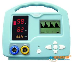
Small handheld oximeters stop there. But larger models, such as the multi-purpose patient monitors used by medics and at hospital bedsides, will also display a waveform. This is a graphical display of the pulsatile flow, with time plotted on the horizontal axis and strength of the detected pulse on the vertical. With a strong, regular pulse, this waveform should be clear and regular, usually with peaked, jagged, or saw-tooth waves. Very small irregular waves, or a waveform with a great deal of artifact, is an indicator that the oximeter is getting a weak signal, and the calculated SpO2 (as well as the calculated pulse) may not be accurate. This waveform can also be used as a kind of “ghetto Doppler,” to help look for the presence of any pulsatile flow in extremities where pulses are not readily palpable. (To be technical, this waveform is known as a photoplethysmograph, or “pleth” for short, and potentially has other applications too– but we’ll leave it alone for now.)

Most modern oximeters, properly functioning and calibrated, have an accuracy between 1% and 2% — call it 1.5% on average. However, their accuracy falls as the saturation falls, and it is generally felt that at saturations below 70% or so, the oximeter ceases to provide reliable readings. Since sats below 90% or so correspond to the “steep” portion of the oxyhemoglobin dissociation curve, where small PaO2 changes might correspond to large changes in SpO2 — in other words, an alarming change in oxygenation status — the fact that your oximeter is losing accuracy in the ranges where you most rely on it is something to keep in mind if using oximetry for continuous monitoring.

The lag time between a change in respiratory conditions (such as increasing supplemental O2 or changing the ventilatory rate) and fully registering this change on the oximeter is usually around 1 minute. And at any given time, the displayed SpO2 is a value calculated by averaging the signal over several seconds, so any near-instantaneous changes should be considered false readings.

Keep reading for our next installment, when we discuss the clinical application of oximetry, and understanding false readings.