Murder by Checklist

Reader Steve Carroll passed along this recent case report from the Annals of Emergency Medicine.

It’s behind a paywall, so let’s summarize.


What happened

A young adult male was shot three times — right lower quadrant, left flank, and proximal right thigh. Both internal and external bleeding were severe. A physician bystander* tried to control it with direct pressure, to no avail.

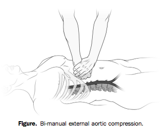
With two hands and a lot of force, however (he weighed over 200 pounds), he was able to hold continuous, direct pressure to the upper abdomen, tamponading the aorta proximal to all three wounds.


Manual aortic pressure


Bleeding was arrested and the patient regained consciousness as long as compression was held. The bystander tried to pass the job off to another, smaller person, who was unable to provide adequate pressure.

When the scene was secured and paramedics arrived, they took over the task of aortic compression. But every time they interrupted pressure to move him to the stretcher or into the ambulance, the patient lost consciousness again. Finally en route, “it was abandoned to obtain vital signs, intravenous access, and a cervical collar.”

The result?

Within minutes, the patient again bled externally and became unresponsive. Four minutes into the 9-minute transfer, he had a pulseless electrical activity cardiac arrest, presumed a result of severe hypovolemia. Advanced cardiac life support resuscitation was initiated and continued for the remaining 5-minute transfer to the ED.

The patient did not survive.


When the cookbook goes bad

The idea of aortic compression is fascinating, but I don’t think it’s the most important lesson to this story.

Much has been said about the drawbacks of rigidly prescriptive protocol-based practice in EMS. But one could argue that our standard teachings allow for you to defer interventions like IV access if you’re caught up preventing hemorrhage. Like they say, sometimes you never get past the ABCs.

The problem here is not necessarily the protocols or the training. It’s the culture. And it’s not just us, because you see similar behavior in the hospital and in other domains.

It’s the idea that certain things just need to be done, regardless of their appropriateness for the patient. It’s the idea that certain patients come with a checklist of actions that need to be dealt with before you arrive at the ED. Doesn’t matter when. Doesn’t matter if they matter.

It’s this reasoning: “If I deliver a trauma patient without a collar, vital signs, and two large-bore IVs, the ER is going to tear me a new one.”

In other words, if you don’t get through the checklist, that’s your fault. But if the patient dies, that’s nobody’s fault.

From the outside, this doesn’t make much sense, because it has nothing to do with the patient’s pathology and what might help them. It has everything to do with the relationship between the paramedic and the ER, or the paramedic and the CQI staff, or the paramedic and the regional medical direction.

Because we work alone out there, without anybody directly overseeing our practice, the only time our actions are judged is when we drop off the patient. Which has led many of us to prioritize the appearance of “the package.” Not the care we deliver on scene or en route. Just the way things look when we arrive.

That’s why crews have idled in ED ambulance bays trying over and over to “get the tube” before unloading. That’s why we’ve had patients walk to the ambulance, climb inside, and sit down, only to be strapped down to a board.

And that’s why we’ve let people bleed to death while we record their blood pressure and needle a vein.

It’s okay to do our ritual checklist-driven dance for the routine patients, because that’s what checklists are for; all the little things that seem like a good idea when there’s time and resources to achieve them. But there’s something deeply wrong when you turn away from something critical — something lifesaving — something that actually helps — in order to achieve some bullshit that doesn’t matter one bit.

If you stop tamponading a wound to place a cervical collar, that cervical collar killed the patient. If you stop chest compressions to intubate, that tube killed the patient. If you delay transport in penetrating trauma to find an IV, that IV killed the patient.

No, let’s be honest. If you do those things, you killed the patient.

Do what actually matters for the patient in front of you. Nobody will ever criticize you for it, and if they do, they are not someone whose criticism should bother you. The only thing that should bother you is killing people while you finish your checklist.


* Correction: the bystander who intervened was not a physician, but “MD” (Matthew Douma), the lead author, who is an RN. — Editor, 7/22/14
Staying in Place: Compensation and Endpoints

Red queen running


Man’s leaning against a wall. He doesn’t move for hours. Just stands there not moving. Finally, someone says, “You been here all day — don’t you have anything to do?”

“I’m doing it,” he answers.

“Doing what?”

“Holding up the wall.”


And who’s to say he’s not? Maybe he’s working as hard as he can to make sure that wall doesn’t fall down.

In this situation, the man is a compensating mechanism. He is struggling to prevent changes in the wall; keeping that wall upright is an endpoint he cares to maintain, to sustain, to keep intact.

How do we know that the wall isn’t holding up the man? Because we don’t care about the man. Whether he leans or falls doesn’t matter much to anybody. But it would be a terrible thing if the wall collapsed. So we’ll let the man lean or shift in order to prop up the wall when it starts to totter — we’ll use him, adjust him, to compensate for any wall-changes. That’s why he’s there.

If the wall gets weak enough or tilts too far, though, he won’t be able to keep it up. He’ll try, but he’s not infinitely strong, and then maybe the wall begins to tilt or collapses completely. Since we know that under normal circumstances, he’s doing his best to prevent this, if we walk in and see that the wall is tilting, that is not a good sign. It may mean that despite his best efforts, the man has exhausted his strength and is no longer able to resist further wall-changes; or it may mean that, for some reason, the man isn’t doing his job properly. Either way, any further tilting will be unopposed, and will probably happen rapidly and uncontrollably.


Compensators and endpoints

This same dynamic plays out within the human body. As we know, living organisms seek to maintain a certain homeostatic equilibrium. We put our vital metabolic processes in motion and we don’t want them to halt or change, despite any insults or fluctuations imposed upon us by our surrounding environment. So our bodies struggle to keep all of our complex systems at an even keel, using a diverse and powerful array of knobs, dials, and other regulatory tools. Not too hot or too cool, not too acid or too basic, not too fast or too slow. Just right.

The kicker is this, however. Some of our physical parameters are more important than others. In other words, while some parameters have room to adjust, others aren’t negotiable, can’t change much, without derailing our basic ability to function and survive. Things like blood pressure (or at least tissue perfusion, for which blood pressure is a pretty good surrogate measure) are essential to life; your pressure can fluctuate a little, but if it drops too low, you are unquestionably going to suffer organ damage and then die. And yet there are many insults that could potentially lower our blood pressure if we let them: if we bleed a little, or pee a little, or don’t drink enough water, or sweat, or even just stand up instead of sitting down. How do we preserve this vital parameter despite such influences?

By compensating, of course. Our body gladly modulates certain processes in order to preserve other, more important parameters. So in order to maintain blood pressure, perhaps we accelerate our heartrate. In an ideal world, it might be nice if the heart were thumping along at — let’s say — a mellow 80 beats per minute. It’ll use little less energy and less oxygen than if it were beating faster. But it’s really important to keep our blood pressure up, and speeding up the heart can increase the pressure, so we gladly make that trade and induce tachycardia. (Many of these compensatory systems are linked to the sympathetic nervous system, our body’s standard “all hands on deck” response to stress and crisis.)

So imagine we find a patient who’s bleeding and notice that he’s tachycardic, with a normal blood pressure. This suggests a compensated shock; the body is using tachycardia to maintain that normal pressure we see; although his volume is lower than usual, the critical endpoint of adequate blood pressure is still intact.

But what if instead, we found him tachycardic and hypotensive? Well, that’s not good. We see that the body is trying to compensate, but we also see that the important endpoint — blood pressure — is falling nonetheless. The body would never intentionally allow that; BP is too important. So we recognize this as decompensated shock. The hypovolemia has progressed so far, and volume is now so low, that he can’t make up the difference anymore — the compensatory slack has run out — and any further decreases in volume will probably lead to an immediate and unopposed drop in pressure. There’s nothing more the body can do on its own; it’s out of rope.

The skilled clinician — or “homeostatic technician” as Jeff Guy says — uses this predictable progression to understand what’s happening in almost any crisis. Because primary insults are initially covered up by compensatory mechanisms, they may not be immediately apparent, and the earliest and most detectable signs of physical insult are usually nothing more than the footprints of the answering compensation. Thus, when when we encounter those, we know to suspect the underlying problem even if it’s not obvious yet. It’s like seeing brakelights flash from cars on the road ahead; even if you can’t see an obstacle yet, you know people are slowing down for something.

Obvious signs of decompensation usually show up late. Once the primary, underlying problem is revealed by failure of the corrective mechanisms, it’s often progressed so far that it’s too late to address. If you wait to brake until you can see the wreck itself, you might not be able to stop in time.


Two signposts for decompensation

There are two great ways to recognize which signs and symptoms connote decompensation.

The first is to understand which physical parameters are endpoints — which functions the body tries to preserve at all costs. These processes are only compromised as a last resort, so if you see them deteriorate, things are in the end-game; the body doesn’t intentionally sacrifice these for the benefit of anything else.

The second clue is more subtle. In this case, you observe a compensatory mechanism (not an endpoint), but find that it’s no longer successfully compensating — it’s failing, and starting to unwind and scale back, rather than doing its job. The changes in the compensatory system are inappropriate, resulting in less of what we need, not more. This happens when our systems are so damaged that they can’t even fix problems and pursue homeostasis anymore; our infrastructure, maintenance, and repair systems are breaking down. Consider this: we saw how tachycardia could be compensatory, but could bradycardia ever be beneficial in shock? Probably not. So if we found a shocked patient with bradycardia (and likely hypotension, the failing endpoint), we should be very alarmed indeed. There’s nothing helpful, compensatory, or beneficial about bradycardia in the setting of shock, so we recognize that the body would never go there on purpose. It’ll only happen when the machinery itself is falling apart.

Consider, for instance, Cushing’s Triad, the collection of signs often encountered after severe traumatic brain injury, when intracranial pressure has increased enough to squeeze the brain out from the skull like toothpaste. The triad includes hypertension, bradycardia, and irregular or slow respirations. What’s interesting is that, while all are a result of increased ICP, one of these is compensatory, while the others are merely the result of damage. Hypertension is the body’s compensatory attempt to force blood into the brain despite the elevated pressure in the skull. But bradycardia and bradypnea simply result from pressure upon the regulatory centers of the brain tasked with maintaining breathing and heart-rate. That’s why hypertension may be seen earlier, while the other two signs won’t usually manifest until the brain is actively herniating. One signals compensation, the other two decompensation.

Of course, there can be other reasons why compensatory mechanisms might fail, or at least exhibit lackluster performance. Some medications or other aspects of a medical history (potentially unrelated to the current complaint) might throw a wrench in the system. For instance, beta blockers (such as metoprolol and other -olol drugs) limit heart-rate as part of their basic mechanism, so patients with beta blockade often have trouble mustering compensatory tachycardia during shock states. That doesn’t mean they’re any less shocked; in fact, it means they’re more susceptible to hypotension, and that you must be especially on the lookout, because you won’t see one of the red flags (a rapid heart-rate) you might usually expect. Elderly patients with many comorbidities are generally not able to muster up effective compensation for anything, so they can deteriorate quickly, and without much fanfare. Ironically, healthy pediatric patients are the opposite: since they’re so “springy” and smoothly functioning, they compensate very well, with few changes in observable endpoints, until suddenly running out of slack and crashing hard because they’re already so far from shore.

Here are a few important compensatory signs, breakdowns of compensatory systems, and vital physical endpoints:


Appropriate signs of compensation

  • Tachycardia — increases cardiac output
  • Vasoconstriction (cool, pale skin) — raises blood pressure
  • Diaphoresis (sweatiness) — decreases temperature when necessary, but is often just a side effect of sympathetic stimulation
  • Tachypnea — increases oxygenation, CO2 blowoff, and cardiac preload
  • Fever — part of the immune system’s response to infection
  • Shivering — warms a hypothermic body

Inappropriate changes in compensatory mechanisms

  • Bradycardia — reduces cardiac output, rarely useful in illness; as a chronic finding may be the result of high levels of cardiovascular fitness (in healthy young patients) or medications (in sick old patients); but acutely, it is an ominous finding
  • Bradypnea — reduces oxygenation, CO2 blowoff, and cardiac preload
  • Hypothermia (or normothermia when a fever is expected) — suggests a failure of temperature regulation

Inviolable endpoints

  • Blood pressure — can elevate in stress states, but should not drop below resting levels
  • Mental status — except in the presence of a drug or similar agent directly affecting cognition, maintaining appropriate alertness and mentation are always a top priority for the body
  • Blood glucose — kept at normal levels in almost all situations, except when the regulatory systems fail, as in diabetes mellitus
  • pH — most of the cellular machinery fall apart if significant acidosis or alkalosis occurs
  • Low O2 saturation or cyanosis — although oxygen saturation can dip briefly without harm, and in some patients (particularly those with COPD, or long-time smokers) it may run low at baseline, a significant acute drop — or the clinical equivalent, which is frank cyanosis — is always inappropriate.

Cuff Links and Hijinks

Any decent EMT can take the austere equipment he’s got and use it to craft all manner of weird and wonderful solutions for the challenges of prehospital medicine. Of course, doing this means understanding the tools you’ve got and all of their powers. Here are a few ideas for using the ubiquitous blood pressure cuff or sphygmomanometer. (We’ve mentioned many of these in passing before, but it’s nice to see them in living color.)


Calibrating the gauge

How to use a pair of pliers to zero the needle on a mis-calibrated dial.


Measuring airway pressure, tourniquets, and cushions

Three handy tricks: first, a method of repurposing common items to create a BVM that provides real-time measurement of the pressure created during positive-pressure ventilation (a very handy teaching tool). Second, using the BP cuff as a tourniquet. Third, using it as an air pillow to fill voids during spinal immobilization.


Do you have a trick for the blood pressure cuff we haven’t mentioned? Let’s hear it!

Understanding Shock IX: Assessment and Recognition

To wrap up our story on shock, let’s discuss how to recognize it.

We all have some idea what shock looks like. Like many pathologies, its loudest early markers are actually indirect — we’ll often recognize the body’s reactions to shock rather than the shock itself.

Although there are a few ways to classify the stages of shock, let’s just use three categories here.


Early or Insignificant

Shock that is very early or minimal in effect may have no particular manifestations. One situation where significant or late shock may also be “hidden” is in the elderly patient, or anyone with significant comorbidities; if their body’s ability to mobilize its compensatory mechanisms is poor, then the red flags won’t be as obvious. This doesn’t mean the shock isn’t as bad; in fact, it means that it’s worse, because their body can’t do as much to mitigate it.

The way to recognize shock at this stage is from the history. If we see an obvious bullet hole in the patient’s chest, and three liters of blood pooling on the ground beside him, then it doesn’t matter how the patient presents otherwise; we’re going to assume that shock is a concern. Blood volume is proportional to bodyweight, but for a typical adult, a fair rule of thumb is to assume about 5-7 liters of total volume. (Not sure what a liter looks like? The bags of saline the medics usually carry are a liter; so are those Nalgene water bottles many people drink from. “Party size” soda bottles are two liters.) Losing more than a liter or two rapidly is difficult to compensate for.

Remember, of course, that blood can also be lost internally, and aside from the occasional pelvic fracture or hemothorax, the best environment for this is the abdomen. Always examine and palpate the abdomen of the trauma patient, looking for rigidity, tenderness, or distention. Remember also that the GI tract is a great place to lose blood; be sure to ask your medical patients about blood or “coffee grounds” (old blood) in the vomit or stool.

Fluid enters and leaves the body continuously, and any disruption in this should be recognized. If a patient complains “I haven’t been able to eat or drink anything in two days,” they’re telling you that they haven’t taken in any fluid for 48 hours. If they tell you they’ve been vomiting or experiencing profuse diarrhea, that’s fluid leaving their body in significant volumes. What about the man who just ran a marathon and sweated out a gallon? Did he drink a gallon to replace it?


Compensated Shock

Significant shock will result in the body attempting to compensate for the low blood volume. Much of this work is done by the sympathetic system, and there are two primary effects: vasoconstriction and cardiac stimulation.

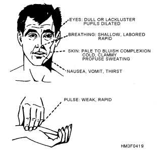
By constricting the blood vessels, we can maintain a reasonable blood pressure and adequate flow even with a smaller circulating volume. We normally vasoconstrict in the periphery — particularly the outer extremities and skin — “stealing” blood from those less-important tissues and retaining it in the vital core. This causes pallor (paleness) and coolness of the external skin. The sympathetic stimulation may also cause diaphoresis (sweating), which is not compensatory, but simply a side effect of the adrenergic release.

The heart also kicks into overdrive, trying to keep the remaining volume moving faster to make up for the loss. It beats faster (chronotropy) and harder (inotropy), resulting in tachycardia. Note that patients who use beta blockers (such as metoprolol) may not be able to muster much, if any, compensatory tachycardia.

A narrowing pulse pressure (the difference between the systolic and diastolic numbers) may be noted; since the diastolic reflects baseline pressure and the systolic reflects the added pressure created by the pumping of the heart, a narrow pulse pressure suggests that cardiac output is diminishing (due to loss of preload), and that more and more of the pressure we’re seeing is simply produced by shrinking the vasculature.

Tachypnea (rapid respirations) are also typically seen. In some cases, this may be due to emotional excitement, and there is also a longstanding belief that it reflects the body’s attempts to “blow off” carbon dioxide and reduce the acidosis created by anaerobic metabolism. (Interestingly, lactate — a byproduct of anaerobic metabolism — can be measured by lab tests, and is also a sign of shock, particularly useful in sepsis.) Additionally, it ensures that all remaining blood has the greatest possible oxygenation. However, it is also plausible that this tachypnea serves to assist the circulatory system: by creating negative pressure in the thorax (the “suction” you make in your chest whenever you inhale) and positive pressure in the abdomen (due to the diaphragm dropping down), you “milk” the vena cava upward during inspiration, improving venous return to the heart and allowing greater cardiac output. This “bellows” effect helps the heart fill more and expel more with each beat.

The more functional the patient’s body is — such as the young, strong, healthy victim — the more effective these compensatory systems will be. Hence the old truism that pediatric patients “fall off a cliff” — they may look great even up through quite profound levels of shock, due to their excellent ability to compensate, then when they finally run out of room they’re already so far in the hole that they become rapidly unhinged. It’s great that these people can compensate well, but it does mean we need to have a high index of suspicion, looking closely for signs of compensation (such as tachycardia) rather than outright signs of shock — because by the time the latter appears, it may be very late indeed.

Patients in compensated shock may become orthostatic; their bodies are capable of perfusing well in more horizontal postures, but when gravity pulls their remaining blood away from the core, this added challenge makes the hypovolemia noticeable. Less acute shock due to causes like dehydration may result in dry skin (particularly the mucus membranes; try examining the inside of the lower eyelid) with poor turgor (pinch a “tent” out of their skin and release it; does it snap back quickly or sluggishly?), and potentially with complaints of thirst. Urine output will usually be minimal. Generally, the more gradually the hypovolemia sets in, the more gradually it can be safely corrected; it’s the sudden, acute losses from causes like bleeding that we’re most worried about.


Decompensated Shock

As shock continues, compensatory systems will struggle harder and harder to maintain perfusion and pressure. Eventually they will fail; further vasoconstriction will reduce rather than improve organ perfusion, beating the heart faster will expel less rather than more blood, and the blood pressure will start to drop.

The hallmark of this stage of shock is the normal functioning of the body beginning to fail. The measured blood pressure will decrease and eventually become unobtainable. Pulses will weaken until they cannot be palpated. As perfusion to the brain decreases, the patient’s mental status will deteriorate. Heart rate and respirations, previously rapid, will begin to slow as the body loses the ability to drive them; like a government office that can’t pay its workers, the regulatory systems that should be fighting the problem begin to shutter their own operations. As the heart continues to “brady down,” eventually it may lose coherence (ventricular fibrillation), or keep stoically trying to contract until the last, but lose all effective output due to the lack of available blood (PEA). Cardiac arrest ensues, with dismal chances for resuscitation.


Alternative Forms of Shock

Although we have focused so far on hypovolemic shock, particularly of traumatic etiology, there are other possibilities. A wide range of shock types exist, but speaking broadly, there are only two other categories important to us: distributive, and cardiogenic/obstructive.

Distributive shocks include anaphylactic, septic, and neurogenic. The essential difference here is that rather than any loss of fluid, the vasculature has simply expanded. Rather than squeezing down on the blood volume to maintain an appropriate pressure, the veins and arteries have gone “slack,” and control of the circulating volume has been lost; it’s simply puddled, like standing water in a sewer pipe. (Depending on the type of shock there may also be some true fluid losses due to edema and third-spacing.) Imagine tying your shoes: in order to stay securely on your feet, the laces need to be pulled snugly (not too tight, not too loose). If the knot comes undone and the laces lose their tension, the shoe will likely slip right off. Your foot hasn’t gotten smaller, but the shoe needs to be hugging it properly to stay in place, and it’s no longer doing its job.

The hallmark of distributive shock is hyperemic (flush or highly perfused) rather than constricted peripheral circulation. The visible skin is warm (or hot) and pink (or red), and the patient may be profoundly orthostatic. Septic shock is associated with infection; anaphylactic with an allergic trigger; and neurogenic with an injury to the spinal cord.

Cardiogenic and obstructive shocks are a different story. In this case, there’s nothing wrong with the circulating volume, or with the vasculature it flows within; instead, there’s a problem with the pump. Cardiogenic shock typically refers to situations like a post-MI heart that’s no longer pumping effectively. Obstructive shock refers to the special cases of pericardial tamponade, massive pulmonary embolism, or tension pneumothorax: physical forces are preventing the heart from expanding or blood from entering it, and hence (despite an otherwise functional myocardium) it’s unable to pump anything out. In either case, we can expect a clinical picture generally similar to hypovolemic shock, but likely with cardiac irregularities — such as ischemic changes or loss of QRS amplitude on the ECG, irregularity or slowing of the pulse, or changes in heart tone (such as muffling) upon auscultation. Pulsus paradoxus (a drop in blood pressure — usually detected by the strength of the palpable pulses — during the inspiratory phase of breathing), electrical alternans (alternating QRS amplitudes on the ECG), and jugular vein distention also may be present in the case of tamponade or severe tension pneumothorax.


In sum, remember these general points:

  1. The history and clinical context should be enough to make you suspect shock even without other signs or symptoms.
  2. The faster the onset, the more urgent the situation; acute shock needs acute care.
  3. Look both for signs of compensation (such as tachycardia) and for signs of decompensation (such as falling blood pressure). However, remember that due to confounding factors (such as particularly effective or ineffective compensatory ability, or pharmacological beta blockade), any or all of these may be absent.
  4. Distributive shocks are mainly characterized by well-perfused peripheral skin; cardiogenic/obstructive shocks are characterized by cardiac irregularities.

Interested parties can stay tuned for a brief appendix discussing fluid choices for resuscitation — otherwise, this journey through shock is finally finished!


Go to Part X (appendix) or back to Part VIII

Understanding Shock VII: Negatives of Fluid Resuscitation

The last time we talked, we learned about the arguments in favor of non-blood fluid resuscitation. What are the arguments against it?


The “blow out the clots” argument

The vascular system is a pressurized circuit. Bleeding means poking an opening in this circuit, and we know that repairing this hole is our number one priority.

The body is pretty good at fixing leaks in its vasculature. But it’s not magic. It’s going to try to form a stable clot that covers and seals the hole, just like wrapping tape around a leaky pipe fitting.

What’s a good way to make this task harder? Increase the pressure inside the pipe. The faster that blood wants to rush out of the hole, the tougher it’s going to be to get a clot to stick there.

Imagine your inflatable raft has a pinhole in it, so you cover it with a piece of tape. It seals well. Then you drop a cooler of beer onto the raft, increasing the internal pressure. The tape blows off. Simple.

Many providers have therefore moved towards the practice of permissive hypotension — resuscitating only to a lower than normal blood pressure — and/or delayed resuscitation — waiting for substantial fluid replacement until bleeding has been controlled. Permissive may mean a pressure of 80, 90, or 100; it may mean giving crystalloids sparingly and only until blood becomes available; or it may mean giving nothing at all except the good stuff. Or you can take a page from the military, which says to resuscitate until a radial pulse is palpable, and the patient’s mental status is restored — then stop.


The dilution argument

There’s another reason why filling the patient with salt water might make it harder to control their bleeding.

Their body is trying to build clots at the location of injury. We want to encourage this process. In order to occur, it requires the activity of circulating platelets and clotting factors.

Mixing the patient’s blood with saline increases its volume but doesn’t increase the number of these clotting precursors. In other words, we’re diluting their blood, just like a bartender watering down your drink. There’s more volume in your cup, but there’s no more of the stuff we care about. And since the ability to form clots is closely related to the concentration of the clotting components, diluting the blood means slower clotting.

Together, these two arguments form a compelling case against the “volume for the sake of volume” theory. The patient’s ability to form clots and stop the bleeding isn’t a small thing; in a way, it’s the only thing. In fact, INR (a measure of clotting speed) has been shown to be a key predictor of whether a trauma patient will survive their injuries.


The proinflammatory argument

One of the key forces in the shock cascade is inflammation. So it seems like promoting more inflammation is the last thing we’d want.

But surprise: infusing fluids can do exactly this. It’s not entirely clear why this happens, but it’s unquestionably true; fluids encourage the inappropriate immune response and increase inflammation and tissue dysfunction. Suffice to say that this is bad.

Back in Vietnam, when aggressive fluid resuscitation really became trendy, doctors were perplexed to find many of their volume-resuscitated patients with a severe condition called “Da Nang lung” (nowadays Acute Respiratory Distress Syndrome) — wet, failing, edematous lungs with no cardiac cause. The combination of increased fluid volume plus increased inflammation means failing lungs. Or check your nearest ICU to see some abdominal compartment syndrome, where fluid fills the abdomen until the organs fail. What were you were saying about fluids being harmless?


The acidosis argument

The pH of our bodies is a hair over 7. Pick up the nearest bag of normal saline and read the label. What’s its pH?

Is it 7? No? More like between 5.0 and 6.0? Interesting. Remember that pH is a logarithmic scale, so we’re talking a difference of 10–100 here. So that nice “normal” fluid can promote significant acidosis.

Is this bad? Only if you like clotting. Acidosis is detrimental to coagulation (among other things), for reasons we’ll get into later. Clotting is good!


The what’s-the-point? argument

In the end, the most compelling argument against pouring what amounts to water into trauma patients is this: fundamentally it is not what they need. Their problem is not a lack of normal saline. “When I find a patient who’s bleeding crystalloid,” some providers are fond of saying, “I’ll give them crystalloid. But usually, the puddle on the ground is blood.”

Now, in some patients, crystalloid may indeed be what’s missing; we’ll touch upon situations like sepsis and dehydration later. But if they’re bleeding, it seems like — at best — playing with any fluid except those that can restore oxygen-carrying capacity or promote clotting is a waste of time that could be spent patching the hole and rushing toward surgery. And at worst, it may be exacerbating the problem.

For a long time, paramedics were taught to fill the hypotensive patient with fluid until their blood pressure was normal. The jury is still out on the best practices for fluid resuscitation, but there is fairly widespread agreement now that this is a bad idea. Many progressive systems have gone the route of giving no crystalloid whatsoever for hemorrhagic shock, or at least giving it very sparingly. Seeing the numbers 120/80 on the monitor seems like a good thing, but shock is not a blood pressure, raising the blood pressure is not necessarily beneficial, and we’re supposed to be making the patient feel better, not ourselves.

So, stop the bleeding, and restore the stuff that matters. Since we rarely give blood in the field, the first one is the main business of EMS. And oddly enough, it’s very much a BLS skill.

Summary:

  1. Increasing the blood pressure interferes with bleeding control.
  2. Diluting the blood discourages clotting while doing nothing for oxygen transport.
  3. Aggressive fluid resuscitation promotes inflammation, edema, and organ dysfunction.
  4. Current best practices are unclear, but likely involve a minor role for crystalloid resuscitation, in favor of bleeding control, blood products, and early surgical intervention.

Next time: mastering the field treatment of hemorrhagic shock.


Go to Part VIII or back to Part VI