Murder by Checklist

Reader Steve Carroll passed along this recent case report from the Annals of Emergency Medicine.

It’s behind a paywall, so let’s summarize.


What happened

A young adult male was shot three times — right lower quadrant, left flank, and proximal right thigh. Both internal and external bleeding were severe. A physician bystander* tried to control it with direct pressure, to no avail.

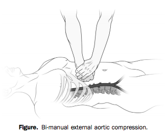
With two hands and a lot of force, however (he weighed over 200 pounds), he was able to hold continuous, direct pressure to the upper abdomen, tamponading the aorta proximal to all three wounds.


Manual aortic pressure


Bleeding was arrested and the patient regained consciousness as long as compression was held. The bystander tried to pass the job off to another, smaller person, who was unable to provide adequate pressure.

When the scene was secured and paramedics arrived, they took over the task of aortic compression. But every time they interrupted pressure to move him to the stretcher or into the ambulance, the patient lost consciousness again. Finally en route, “it was abandoned to obtain vital signs, intravenous access, and a cervical collar.”

The result?

Within minutes, the patient again bled externally and became unresponsive. Four minutes into the 9-minute transfer, he had a pulseless electrical activity cardiac arrest, presumed a result of severe hypovolemia. Advanced cardiac life support resuscitation was initiated and continued for the remaining 5-minute transfer to the ED.

The patient did not survive.


When the cookbook goes bad

The idea of aortic compression is fascinating, but I don’t think it’s the most important lesson to this story.

Much has been said about the drawbacks of rigidly prescriptive protocol-based practice in EMS. But one could argue that our standard teachings allow for you to defer interventions like IV access if you’re caught up preventing hemorrhage. Like they say, sometimes you never get past the ABCs.

The problem here is not necessarily the protocols or the training. It’s the culture. And it’s not just us, because you see similar behavior in the hospital and in other domains.

It’s the idea that certain things just need to be done, regardless of their appropriateness for the patient. It’s the idea that certain patients come with a checklist of actions that need to be dealt with before you arrive at the ED. Doesn’t matter when. Doesn’t matter if they matter.

It’s this reasoning: “If I deliver a trauma patient without a collar, vital signs, and two large-bore IVs, the ER is going to tear me a new one.”

In other words, if you don’t get through the checklist, that’s your fault. But if the patient dies, that’s nobody’s fault.

From the outside, this doesn’t make much sense, because it has nothing to do with the patient’s pathology and what might help them. It has everything to do with the relationship between the paramedic and the ER, or the paramedic and the CQI staff, or the paramedic and the regional medical direction.

Because we work alone out there, without anybody directly overseeing our practice, the only time our actions are judged is when we drop off the patient. Which has led many of us to prioritize the appearance of “the package.” Not the care we deliver on scene or en route. Just the way things look when we arrive.

That’s why crews have idled in ED ambulance bays trying over and over to “get the tube” before unloading. That’s why we’ve had patients walk to the ambulance, climb inside, and sit down, only to be strapped down to a board.

And that’s why we’ve let people bleed to death while we record their blood pressure and needle a vein.

It’s okay to do our ritual checklist-driven dance for the routine patients, because that’s what checklists are for; all the little things that seem like a good idea when there’s time and resources to achieve them. But there’s something deeply wrong when you turn away from something critical — something lifesaving — something that actually helps — in order to achieve some bullshit that doesn’t matter one bit.

If you stop tamponading a wound to place a cervical collar, that cervical collar killed the patient. If you stop chest compressions to intubate, that tube killed the patient. If you delay transport in penetrating trauma to find an IV, that IV killed the patient.

No, let’s be honest. If you do those things, you killed the patient.

Do what actually matters for the patient in front of you. Nobody will ever criticize you for it, and if they do, they are not someone whose criticism should bother you. The only thing that should bother you is killing people while you finish your checklist.


* Correction: the bystander who intervened was not a physician, but “MD” (Matthew Douma), the lead author, who is an RN. — Editor, 7/22/14
Staying in Place: Compensation and Endpoints

Red queen running


Man’s leaning against a wall. He doesn’t move for hours. Just stands there not moving. Finally, someone says, “You been here all day — don’t you have anything to do?”

“I’m doing it,” he answers.

“Doing what?”

“Holding up the wall.”


And who’s to say he’s not? Maybe he’s working as hard as he can to make sure that wall doesn’t fall down.

In this situation, the man is a compensating mechanism. He is struggling to prevent changes in the wall; keeping that wall upright is an endpoint he cares to maintain, to sustain, to keep intact.

How do we know that the wall isn’t holding up the man? Because we don’t care about the man. Whether he leans or falls doesn’t matter much to anybody. But it would be a terrible thing if the wall collapsed. So we’ll let the man lean or shift in order to prop up the wall when it starts to totter — we’ll use him, adjust him, to compensate for any wall-changes. That’s why he’s there.

If the wall gets weak enough or tilts too far, though, he won’t be able to keep it up. He’ll try, but he’s not infinitely strong, and then maybe the wall begins to tilt or collapses completely. Since we know that under normal circumstances, he’s doing his best to prevent this, if we walk in and see that the wall is tilting, that is not a good sign. It may mean that despite his best efforts, the man has exhausted his strength and is no longer able to resist further wall-changes; or it may mean that, for some reason, the man isn’t doing his job properly. Either way, any further tilting will be unopposed, and will probably happen rapidly and uncontrollably.


Compensators and endpoints

This same dynamic plays out within the human body. As we know, living organisms seek to maintain a certain homeostatic equilibrium. We put our vital metabolic processes in motion and we don’t want them to halt or change, despite any insults or fluctuations imposed upon us by our surrounding environment. So our bodies struggle to keep all of our complex systems at an even keel, using a diverse and powerful array of knobs, dials, and other regulatory tools. Not too hot or too cool, not too acid or too basic, not too fast or too slow. Just right.

The kicker is this, however. Some of our physical parameters are more important than others. In other words, while some parameters have room to adjust, others aren’t negotiable, can’t change much, without derailing our basic ability to function and survive. Things like blood pressure (or at least tissue perfusion, for which blood pressure is a pretty good surrogate measure) are essential to life; your pressure can fluctuate a little, but if it drops too low, you are unquestionably going to suffer organ damage and then die. And yet there are many insults that could potentially lower our blood pressure if we let them: if we bleed a little, or pee a little, or don’t drink enough water, or sweat, or even just stand up instead of sitting down. How do we preserve this vital parameter despite such influences?

By compensating, of course. Our body gladly modulates certain processes in order to preserve other, more important parameters. So in order to maintain blood pressure, perhaps we accelerate our heartrate. In an ideal world, it might be nice if the heart were thumping along at — let’s say — a mellow 80 beats per minute. It’ll use little less energy and less oxygen than if it were beating faster. But it’s really important to keep our blood pressure up, and speeding up the heart can increase the pressure, so we gladly make that trade and induce tachycardia. (Many of these compensatory systems are linked to the sympathetic nervous system, our body’s standard “all hands on deck” response to stress and crisis.)

So imagine we find a patient who’s bleeding and notice that he’s tachycardic, with a normal blood pressure. This suggests a compensated shock; the body is using tachycardia to maintain that normal pressure we see; although his volume is lower than usual, the critical endpoint of adequate blood pressure is still intact.

But what if instead, we found him tachycardic and hypotensive? Well, that’s not good. We see that the body is trying to compensate, but we also see that the important endpoint — blood pressure — is falling nonetheless. The body would never intentionally allow that; BP is too important. So we recognize this as decompensated shock. The hypovolemia has progressed so far, and volume is now so low, that he can’t make up the difference anymore — the compensatory slack has run out — and any further decreases in volume will probably lead to an immediate and unopposed drop in pressure. There’s nothing more the body can do on its own; it’s out of rope.

The skilled clinician — or “homeostatic technician” as Jeff Guy says — uses this predictable progression to understand what’s happening in almost any crisis. Because primary insults are initially covered up by compensatory mechanisms, they may not be immediately apparent, and the earliest and most detectable signs of physical insult are usually nothing more than the footprints of the answering compensation. Thus, when when we encounter those, we know to suspect the underlying problem even if it’s not obvious yet. It’s like seeing brakelights flash from cars on the road ahead; even if you can’t see an obstacle yet, you know people are slowing down for something.

Obvious signs of decompensation usually show up late. Once the primary, underlying problem is revealed by failure of the corrective mechanisms, it’s often progressed so far that it’s too late to address. If you wait to brake until you can see the wreck itself, you might not be able to stop in time.


Two signposts for decompensation

There are two great ways to recognize which signs and symptoms connote decompensation.

The first is to understand which physical parameters are endpoints — which functions the body tries to preserve at all costs. These processes are only compromised as a last resort, so if you see them deteriorate, things are in the end-game; the body doesn’t intentionally sacrifice these for the benefit of anything else.

The second clue is more subtle. In this case, you observe a compensatory mechanism (not an endpoint), but find that it’s no longer successfully compensating — it’s failing, and starting to unwind and scale back, rather than doing its job. The changes in the compensatory system are inappropriate, resulting in less of what we need, not more. This happens when our systems are so damaged that they can’t even fix problems and pursue homeostasis anymore; our infrastructure, maintenance, and repair systems are breaking down. Consider this: we saw how tachycardia could be compensatory, but could bradycardia ever be beneficial in shock? Probably not. So if we found a shocked patient with bradycardia (and likely hypotension, the failing endpoint), we should be very alarmed indeed. There’s nothing helpful, compensatory, or beneficial about bradycardia in the setting of shock, so we recognize that the body would never go there on purpose. It’ll only happen when the machinery itself is falling apart.

Consider, for instance, Cushing’s Triad, the collection of signs often encountered after severe traumatic brain injury, when intracranial pressure has increased enough to squeeze the brain out from the skull like toothpaste. The triad includes hypertension, bradycardia, and irregular or slow respirations. What’s interesting is that, while all are a result of increased ICP, one of these is compensatory, while the others are merely the result of damage. Hypertension is the body’s compensatory attempt to force blood into the brain despite the elevated pressure in the skull. But bradycardia and bradypnea simply result from pressure upon the regulatory centers of the brain tasked with maintaining breathing and heart-rate. That’s why hypertension may be seen earlier, while the other two signs won’t usually manifest until the brain is actively herniating. One signals compensation, the other two decompensation.

Of course, there can be other reasons why compensatory mechanisms might fail, or at least exhibit lackluster performance. Some medications or other aspects of a medical history (potentially unrelated to the current complaint) might throw a wrench in the system. For instance, beta blockers (such as metoprolol and other -olol drugs) limit heart-rate as part of their basic mechanism, so patients with beta blockade often have trouble mustering compensatory tachycardia during shock states. That doesn’t mean they’re any less shocked; in fact, it means they’re more susceptible to hypotension, and that you must be especially on the lookout, because you won’t see one of the red flags (a rapid heart-rate) you might usually expect. Elderly patients with many comorbidities are generally not able to muster up effective compensation for anything, so they can deteriorate quickly, and without much fanfare. Ironically, healthy pediatric patients are the opposite: since they’re so “springy” and smoothly functioning, they compensate very well, with few changes in observable endpoints, until suddenly running out of slack and crashing hard because they’re already so far from shore.

Here are a few important compensatory signs, breakdowns of compensatory systems, and vital physical endpoints:


Appropriate signs of compensation

  • Tachycardia — increases cardiac output
  • Vasoconstriction (cool, pale skin) — raises blood pressure
  • Diaphoresis (sweatiness) — decreases temperature when necessary, but is often just a side effect of sympathetic stimulation
  • Tachypnea — increases oxygenation, CO2 blowoff, and cardiac preload
  • Fever — part of the immune system’s response to infection
  • Shivering — warms a hypothermic body

Inappropriate changes in compensatory mechanisms

  • Bradycardia — reduces cardiac output, rarely useful in illness; as a chronic finding may be the result of high levels of cardiovascular fitness (in healthy young patients) or medications (in sick old patients); but acutely, it is an ominous finding
  • Bradypnea — reduces oxygenation, CO2 blowoff, and cardiac preload
  • Hypothermia (or normothermia when a fever is expected) — suggests a failure of temperature regulation

Inviolable endpoints

  • Blood pressure — can elevate in stress states, but should not drop below resting levels
  • Mental status — except in the presence of a drug or similar agent directly affecting cognition, maintaining appropriate alertness and mentation are always a top priority for the body
  • Blood glucose — kept at normal levels in almost all situations, except when the regulatory systems fail, as in diabetes mellitus
  • pH — most of the cellular machinery fall apart if significant acidosis or alkalosis occurs
  • Low O2 saturation or cyanosis — although oxygen saturation can dip briefly without harm, and in some patients (particularly those with COPD, or long-time smokers) it may run low at baseline, a significant acute drop — or the clinical equivalent, which is frank cyanosis — is always inappropriate.

Pulse Oximetry: Application

The final part of a series on oximetry: start with Respiration and Hemoglobin and Pulse Oximetry: Basics

Pulse oximetry is not always available in EMS — depending on level of care, scope of practice in your area, and how your service chooses to equip you — but when it is, it’s a valuable tool in your diagnostic toolbox. Just like we discussed before, and just like any other piece of the patient assessment, using it properly requires understanding how it works and when it doesn’t.


Clinical context: When a sat is not a sat

Simply put, oximetry is the vital sign of oxygenation. It is the direct measurement of the oxygen in your bloodstream. It does not quite measure the oxygen that is actually available to your cells, but it gets close.

First, remember that actual oxygen delivery requires not just adequate hemoglobin saturation, but also enough total hemoglobin, moving around at an adequate rate. In hypovolemia, such as the shocky trauma patient, or in anemia, you might see a high SpO2 — which may be entirely accurate — but this doesn’t necessarily mean that the organs are not hypoxic. After all, you could have nothing but a single lonely hemoglobin floating around, and if it had four oxygen bound to it, you would technically have a sat of 100%. But that won’t keep anyone alive. Evaluating perfusion is a separate matter from evaluating oxygenation.

Second, remember our discussion of the oxyhemoglobin dissociation curve. The fact that you have oxygen bound to your hemoglobin doesn’t mean that it’s actually being delivered to your cells. That is, you can be hypoxic — inadequate cellular oxygenation of your organs — without being hypoxemic — inadequate oxygen present in the blood. Oximetry will only reveal hypoxemia.

Two of the strongest confounders here are cyanide and carbon monoxide (CO) poisoning. The main effect of cyanide is to impair the normal cellular aerobic cycle, preventing the utilization of oxygen; since it has no effect on your lungs or hemoglobin, the result is a normal saturation, yet profound hypoxia, since none of the bound oxygen can actually be used. Carbon monoxide, on the other hand, involves a twofer; it binds to hemoglobin in the place of oxygen, creating a monster called carboxyhemoglobin. CO has far more affinity for carboxyhemoglobin than oxygen does, so it’s hard to dislodge, and you therefore lose 1/4 of your available binding sites in the affected hemoglobin. But it doesn’t stop there. Carboxyhemoglobin also has a higher affinity for oxygen. This creates a leftward shift in the oxyhemoglobin dissociation curve — the oxygen that actually does bind finds itself “stuck,” and these well-saturated boats happily sail past increasingly hypoxic tissues without ever unloading their O2.

Consider the oximetric findings in these patients. The cyanide patient will have unimpaired blood oxygenation, so (unless he has already succumbed to respiratory failure due to the effects), a normal sat will be seen; however, hypoxia will be clinically apparent, particularly as ischemia of the heart and brain. Carbon monoxide, on the other hand, will reveal a normal or elevated (100%) sat which is partially accurate — some of that is true oxygen — and partially baloney, since CO looks the same to the oximeter as O2. But this is moot, because neither the bound CO nor the bound O2 is available to the cells. Oximeters do exist that can detect the presence of carboxyhemoglobin, known as CO-oximeters, but they are expensive and uncommon, and there is some question as to their accuracy. Your best helper here is in the patient history: both CO and cyanide are produced by fires, or any combustion in enclosed spaces (such as stoves or heaters), cyanide being released by the combustion of many plastics. You should be very wary of normal sats in any patient coming from a house fire or similar circumstances.

(Both cyanide and CO poisoning are known for causing bright red skin. In both cases oxygen is not being removed from hemoglobin, so arterial blood remains pink and well-saturated. Carboxyhemoglobin itself is also an unusually bright red. This skin, a late sign, is usually seen in dead or near-dead patients.)

Third, consider that although oximetry is an excellent measure of oxygenation, this is not the same as assessing respiratory status. It’s a little like measuring the blood pressure: although it’s a very important number, BP is an end product of numerous other compensatory mechanisms, and a normal pressure doesn’t mean that there aren’t challenges being placed on it — merely that they’re challenges you’re currently able to compensate for. Perhaps you’re satting 98%, but only by breathing 40 times a minute, and you’re fatiguing fast. Perhaps you’re satting 94%, but your airway is closing quickly and in a few minutes you won’t be breathing at all. These are clinical findings that may not be revealed in SpO2 until it’s too late.

Fourth: oximetry measures oxygenation, but not ventilation. When you breathe in, you inhale oxygen; when you breathe out, you exhale carbon dioxide. Although we use the term ventilation to describe the overall process of breathing, formally in the respiratory world it refers to the removal of carbon dioxide. Is oxygenation the more important of these two functions? Certainly; it will kill you much faster. But hypercapnia (high CO2) caused by inadequate ventilation is also a problem, and pulse oximetry does not measure it. (Capnography is the vital sign of ventilation, but that’s a topic for another day.) Now, insofar as oxygenation is primarily determined by respiratory adequacy (rate, volume, and quality of breathing), and respiration both oxygenates and ventilates, oximetry can be a good indirect measurement of ventilation; if you’re oxygenating well, you’re probably ventilating well too. This remains true if breathing is assisted via BVM, CPAP, or other device. But this is not true if supplemental oxygen is applied. Increasing the fraction of inspired oxygen (FiO2) improves oxygenation without affecting ventilation; on 100% oxygen I might be breathing 8 times a minute, oxygenating well, but ventilating inadequately.

Finally, it’s worth remembering that once you reach 100% saturation, PaO2 may no longer correlate directly with SpO2. If you reach 100% saturation at a PaO2 of 80, we could keep increasing the available oxygen until you hit a PaO2 of 500, but your sat will still read 100%. So without taking a blood gas, we don’t know whether that sat of 100% is incredibly robust, or is very close to desatting. (That’s not to say that a higher PaO2 is necessarily better; recent research continues to suggest that hyperoxygenation is harmful in many conditions. Not knowing the true PaO2 can be problematic in either direction.)


Hardware failure: When a sat is not anything

In what clinical circumstances does oximetry tend to fail? The primary one is when there isn’t sufficient arterial flow to produce a strong signal. This can be systemic, such as hypovolemia — or cardiac arrest — or it can be local, such as in PVD. (The shocked patient has both problems, being both hypovolemic and peripherally vasoconstricted.) Feel the extremity you’re applying the sensor to; if it’s warm, your chances of an accurate reading are good. The best confirmation here is to watch the waveform; a clear, accurate waveform is a very good indicator that you have a strong signal.

Tremors from shivering, Parkinsonism, or fever-induced rigors can also produce artifact on the oximeter. Some patients also just don’t like the probe on their finger. Try holding it in place, keeping the sensor tightly against the skin and the digit motionless. If there’s no luck, try another site. Any finger will work, or any toe, or an earlobe. (Some devices don’t require “sandwiching” the tissue, and can be stuck to the forehead or other proximal site, but these are uncommon in outpatient settings.)

There are a few other situations that can interfere with normal readings. In most cases, nail polish is not a problem, but dark colors do decrease the transmittance, so some shades have been reported to produce falsely low readings in the presence of already low sats or poor perfusion — as always, check your waveform for adequate signal strength. Very bright fluorescent lights have been reported to create strange numbers, and ambient infrared light — such as the heat lamps found in neonatal isolettes — can certainly create spurious readings. A few other medical oddities fall into this category as well, including intravenous dyes like methylene blue, and methemoglobinemia, which produces false sats trending towards 85%.

Is oximetry a replacement for a clinical assessment of respiration, including rate, rhythm, subjective difficulty, breath sounds, skin, and relevant history? Absolutely not. But since none of those actually provide a quantified assessment of oxygenation, they are also no replacement for oximetry. It is a valuable addition to any diagnostic suite, particularly to help in monitoring a patient over time, as well as for detecting depressed respirations before they become clinically obvious — especially in the clinically opaque patient, such as the comatose. When it’s unavailable in the field, we readily do without it. But when it’s available, it’s worth using, and anything worth using is worth understanding.

Pulse Oximetry: Basics

Just tuning in? Start with Respiration and Hemoglobin, or continue to Pulse Oximetry: Application

Once upon a time, the only way to measure SaO2 was to draw a sample of arterial blood and send it down to the lab for a rapid analysis of gaseous contents — an arterial blood gas (ABG), or something similar. This result is definitive, but it takes time, and in some patients by the time you get back your ABG, its results are already long outdated. The invention of a reliable, non-invasive, real-time (or nearly so) method of monitoring arterial oxygen saturation is one of the major advances in patient assessment from the past fifty years.

Oximetry relies on a simple principle: oxygenated blood looks different from deoxygenated blood. We all know this is true. If you cut yourself and bleed from an artery — oxygenated blood — it will appear bright red. Venous blood — deoxygenated — is much darker.

We can take advantage of this. We place a sensor over a piece of your body that is perfused with blood, yet thin enough to shine light through — a finger, a toe, maybe an earlobe. Two lights shine against one side, and two sensors detect this light from the other side. One light is of a wavelength (infared at around 800–1000nm) that is mainly absorbed by oxygenated blood; the other is of a wavelength (visible red at 600–750nm) that is mainly absorbed by deoxygenated blood. By comparing how much of each light reaches the other side, we can determine how much oxygenated vs. deoxygenated blood is present.

The big turning point in this technology came when “oximetry” turned into “pulse oximetry.” See, the trouble with this shining-light trick is that there are a lot of things between light and sensor other than arterial blood — skin, muscle, venous blood, fat, sweat, nail polish, and other things, and all of these might have differing opacity depending on the patient and the sensor location. But what we can do is monitor the amount of light absorbed during systole — while the heart is pumping blood — and monitor the amount absorbed during diastole — while the heart is relaxed — and compare them. The only difference between these values should be the difference caused by the pulsation of arterial blood (since your skin, muscle, venous blood, etc. are not changing between heartbeats), so if we subtract the two, the result should be an absorption reading from SaO2 only. Cool!

Most oximeters give you a few different pieces of information when they’re applied. The most important is the SaO2, a percentage between 0% and 100% describing how saturated the hemoglobin are with oxygen. (Typically, in most cases we refer to this number as SpO2, which is simply SaO2 as determined by pulse oximetry. This can be helpful by reminding us that oximeters aren’t perfect, and aren’t necessarily giving us a direct look at the blood contents, but for most purposes they are interchangeable terms.) But due to the pulse detection we just described, most oximeters will also display a fairly reliable heart rate for you.

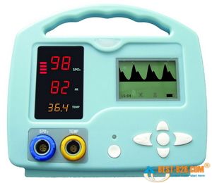
Small handheld oximeters stop there. But larger models, such as the multi-purpose patient monitors used by medics and at hospital bedsides, will also display a waveform. This is a graphical display of the pulsatile flow, with time plotted on the horizontal axis and strength of the detected pulse on the vertical. With a strong, regular pulse, this waveform should be clear and regular, usually with peaked, jagged, or saw-tooth waves. Very small irregular waves, or a waveform with a great deal of artifact, is an indicator that the oximeter is getting a weak signal, and the calculated SpO2 (as well as the calculated pulse) may not be accurate. This waveform can also be used as a kind of “ghetto Doppler,” to help look for the presence of any pulsatile flow in extremities where pulses are not readily palpable. (To be technical, this waveform is known as a photoplethysmograph, or “pleth” for short, and potentially has other applications too– but we’ll leave it alone for now.)

Most modern oximeters, properly functioning and calibrated, have an accuracy between 1% and 2% — call it 1.5% on average. However, their accuracy falls as the saturation falls, and it is generally felt that at saturations below 70% or so, the oximeter ceases to provide reliable readings. Since sats below 90% or so correspond to the “steep” portion of the oxyhemoglobin dissociation curve, where small PaO2 changes might correspond to large changes in SpO2 — in other words, an alarming change in oxygenation status — the fact that your oximeter is losing accuracy in the ranges where you most rely on it is something to keep in mind if using oximetry for continuous monitoring.

The lag time between a change in respiratory conditions (such as increasing supplemental O2 or changing the ventilatory rate) and fully registering this change on the oximeter is usually around 1 minute. And at any given time, the displayed SpO2 is a value calculated by averaging the signal over several seconds, so any near-instantaneous changes should be considered false readings.

Keep reading for our next installment, when we discuss the clinical application of oximetry, and understanding false readings.

Treat the Patient?

We’re taking a short break from our series on transfers to discuss a recent post on the EMT-Medical Student blog. One of the issues he brought up is the old saw, “Treat the patient, not the machine.” Rogue Medic struck on this as well.

What do people mean when they say this? They mean that if you attach a diagnostic tool like a pulse oximeter, and it gives you a result that is at odds with the rest of your assessment, then it is probably wrong, and you should not base your decisions on it. It can be broadened to the BLS level, including findings like vital signs, by saying: “Treat the patient, not the number.”

And it’s essentially true. In fact, something I frequently harp on is that diagnosis must always be based on a broad constellation of consistent findings, not on any one red flag. We like red flags, we want red flags, because they’re easy, but it never works that way. The body is an interdependent system, and if a pathology is present, then it almost always has multiple effects detectable in multiple places.

This idea can be looked at differently by asking another question: is it possible to be severely, acutely sick without showing it? I don’t mean long-term problems like cancer; you can’t look at someone and detect that. But if someone’s dying in front of you, of a proximate cause like hypoxia, is it always obvious based on their presentation?

Generally the answer is yes. That’s why it’s wrongheaded to look at a healthy patient with pink skin, normal respiratory rate, calmly denying shortness of breath, but with a low oxygen saturation, and say, “Oh no — he’s hypoxic!” If your oximeter says 72%, what’s more likely — that the number is wrong, or that the patient is somehow hypoxic without any other evidence of it?

Call this the phenomenon of the Hidden Killer. Is it common? Is it real?

It is not common. But it is real. And that’s what’s not recognized when people say, “Treat the patient…”

Why do we take 12-lead ECGs on chest pain patients? Because a clinical assessment alone cannot reliably detect ST elevation, which (simplifying the issue!) is diagnostic for a heart attack.

Why do we take CT scans of blunt head injury patients? Because a clinical assessment alone cannot reliably detect intracranial hemorrhage.

Why do we perform abdominal ultrasounds in multi-system trauma patients? Because a clinical assessment alone cannot reliably detect abdominal bleeding.

Now, some critics will say that all of these will indeed present with obvious, frank clinical findings. The major STEMI patient will eventually enter cardiogenic shock. The head bleed will become comatose and present with Cushing’s Triad and herniation. The abdominal hemorrhage will have guarding, distension, and eventually outright shock.

All true enough. But we’d like to find them earlier than that. It’s true that severe and late pathologies are usually obvious, but our job is to find them when they can still be treated, not after their effects are permanent or lethal. Heck, we could also just provide no medical care and wait until everyone died to make a diagnosis, which would extremely easy to assess, but a little pointless. It is rare that big problems do not have a big assessment footprint, but “small” problems can still be a big deal.

Consider the much-maligned pulse ox. Surely it does not replace a full assessment. But when used appropriately and its role understood, it provides valuable information. A drop from 99% to 94% saturation may not be clinically obvious, but it is potentially significant and surely worth knowing about. What about the patient who is non-verbal at his baseline? Is he going to complain if he drops from 95% to 87%? Will it be frankly obvious from his skin and breathing? Maybe, maybe not. (How about if he’s on a mechanical ventilator at a fixed rate?) If not obvious, does that mean it’s no big deal?

Is the pulse ox always correct? No. But like all things except magic, it’s wrong in predictable ways, ways that can be accounted for, and when it is wrong, that can tell you something too. It requires adequate peripheral circulation, and poor perfusion will make it read low. How is the patient’s distal perfusion? Pink and warm? Good capillary refill? Then you’re probably okay. Carbon monoxide poisoning will make the sat read high. Has the patient been in enclosed spaces with heaters or open flames? Working around engines? Is there any potential source of CO in their history? If not, you’re probably okay. Alternately, does their sat read unusually high compared to their clinical presentation? You might then consider carbon monoxide — something you might not have otherwise have known without the oximeter. It didn’t give you a correct number, but by knowing how and when it fails, it gave us a useful answer.

Here’s a recent example. I picked up a patient with a blood pressure of 54/4. That is a ridiculous blood pressure; arguably, nobody should have it, on the theory that a pressure that low should be pretty close to unobtainable. But, there it was. We diverted to the nearest hospital and I was subsequently chewed out by the receiving nurse.

Do I think that patient truly had a central arterial pressure of 54/4? Nah. Although she wasn’t doing well, her skin was better than that, and although she was altered and combative, she wasn’t comatose. However, her pressure was undoubtedly low, and just how low? If I don’t go with this number, then I’ve got no guidance. The clinical picture was clouded. I couldn’t ask if she knew what day it was; I couldn’t ask what her complaints were; she was non-verbal. She was tachycardic and hypoxic and diaphoretic; she was certainly sick. So, treat the patient, or treat the number? The number may not have been right, but it was concerning enough that it couldn’t be ignored without an assessment that otherwise screamed “no problems here!”, which was not what we had.

Treat the patient? We always treat the patient. A hands-on physical and history is a vital, vital tool for assessment, but other tools are also useful. Some people lament the downfall of the traditional clinical assessment, from the days when doctors with fingers like pianists made diagnoses from findings like Ewart’s sign, and it is shame, but the reason that the high-tech tools like imaging and labs have become de rigueur is that they work well — they diagnose many problems with a speed, sensitivity, and reliability that is not otherwise possible. Nobody would ever say, “Treat the patient, not the unstable cervical spine fracture,” because we recognize that’s the sort of thing you may not otherwise notice until it’s too late. That’s why we spend big bucks on CT scanners.

It all matters. It’s all useful. We should neither cast aside our individual numbers nor ignore the bigger picture. Data is something that, like money and sex, you can never have too much of.