Understanding Shock VI: Fluid Resuscitation

So we know now that in any hemorrhagic shock, controlling the bleeding is step one, and restoring the supply of something resembling blood is step two. Should we also consider infusing some other fluids, even those that don’t help carry any oxygen?

Why would we even consider such a thing? It would make sense if “fluid” is what we’re missing, which is the case when shock is caused by something like dehydration. But in hemorrhage, we’re missing blood, not water. Still, there are a few reasons this might be worthwhile. Let’s discuss the “pro” arguments first, then come back around and talk about the “cons.”


The hydraulic argument

Fundamentally, the human vascular system is a hydraulic circuit.

In other words, it’s a giant circle of stretchy elastic tubes, like those long circus balloons. It’s all filled with fluid, which stretches out those tubes and pressurizes the whole system. Then a central pump pushes all the fluid in the system around in an endless loop.

One of the properties of such a system is that, without adequate internal pressure, it won’t work. It’s not that it works badly; it just fails altogether. And although pumping harder and faster can help elevate the pressure a little, and squeezing down on the tubes to make them smaller can help more, in the end if there’s not enough fluid in the system, nothing’s moving anywhere. If the heart isn’t filling with a certain amount of blood during diastole, it won’t push it forward during systole; it can’t pump out what it doesn’t take in.

So maybe there’s a certain logic for maintaining an adequate blood pressure, no matter what sort of fluid we’re actually circulating. Although pressure alone doesn’t carry oxygen, maintaining some pressure is certainly a prerequisite for carrying anything. To put it dryly, although BP isn’t everything, people with no BP are dead.

Moreover, some of the pathways in the shock cascade are, perhaps, initiated by low intravascular volume as much as by actual inadequate oxygen delivery. If we can keep the circulating volume pretty decent, maybe we can convince the body that all’s well — no need for a freak-out today.


The extravascular resuscitation argument

Flip back the calendar to the era of the Vietnam War, a landmark time in trauma care. Researchers like Dr. Tom Shires were experimenting on dogs.

They’d do things like drain from them a fixed volume of blood, then clamp off the bleeding and wait for a bit. Then they’d put back every drop of blood they’d removed. Most of the dogs died nonetheless, a phenomenon you and I now understand, since we’re totally experts in the self-sufficiency of the shock process.

But then they’d repeat the experiment. Only this time, rather than just giving the dogs back their blood, they’d also give them some crystalloid fluid. Just water with some stuff like electrolytes in it. This time, more of the dogs survived.

The theory explaining this goes something like so: where is most of the fluid in your body? We know that a high percentage of our bodyweight is water, but does that flow mostly in the blood? Anatomists talk about three different fluid “spaces”: the intravascular space (inside the vessels, where the blood circulates); the intracellular space (the interior of our actual cells); and the interstitial space (the “sea” of fluid permeating the tissue beds but outside the cells, bathing and nourishing them). Fluid moves between these spaces as needed, but at any given time, the majority of your body’s fluid is actually in the interstitial and intracellular (the extravascular) spaces — that is to say, not in the blood at all.

Shock causes increased permeability of the tissues and of the vascular tree, while simultaneously dropping intravascular (hydrostatic) pressure. So when the dogs entered shock, after a short while fluid began to “leak” from the interstitial and intracellular spaces back into the intravascular space. In essence, the dogs’ tissues were returning some of their retained fluid back into the bloodstream — and human tissues do this too. This shift actually increases the vascular volume, which is nice in a sense, and can be seen as a method of compensation: the body is tapping some of its reserve fluid to restore what was lost. However, it does leave the tissues dry. By infusing some saline along with the blood, Shires was helping his test subjects resuscitate both spaces. The intravascular space needed blood, but the extravascular spaces just needed fluid. (Of course, if we replace the blood, eventually the extravascular tissues will be rehydrated and the loaner fluid returned; but if we didn’t provide any extra fluid, that would once again leave the intravascular compartment a little light. Also, some of it — which leaked into neither the intravascular nor extravascular spaces, but the “third space,” areas such as the abdomen where it doesn’t belong — won’t be readily returned at all.)

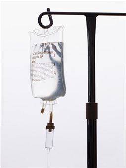
Some combination of these two arguments became the foundation for a decades-long practice whereby hemorrhaging patients are given a certain amount of crystalloid (usually saline, or a modified form of saline like Lactated Ringer’s), often prior or in addition to giving blood products. In many cases this fluid is titrated to maintain a desired blood pressure, and this practice is still widespread today, especially in the prehospital world. In some cases, colloidal fluids (which contain large molecules such as proteins) are also used and have generally similar effects.

Key points:

  1. Bleeding control and restoring actual oxygen-carrying capacity are the main priorities in hemorrhagic shock, but there may also be value in non-blood fluid resuscitation.
  2. One argument for this is the maintenance of adequate blood pressure in order for the circulatory system to function.
  3. Another argument is the replenishment of the fluid lost from extravascular spaces.

Next episode we’ll discuss the dark side of crystalloid resuscitation.
Go to Part VII or back to Part V

Understanding Shock V: Blood Transfusion

So let’s say we’ve stopped the bleeding as best we can. Now what?

The patient is still low on blood, and we know about all the problems this will cause. So shouldn’t we try and give them some back?

Well, maybe.

It makes sense that someone who loses blood should get some blood replaced. And this is a very old concept. Once upon a time, we simply drew blood from one person and gave it to another — a process that was greatly improved when we learned how to screen and test blood for compatibility and disease. This method is still used in some settings, such as the military, which treats its entire force as a “walking blood bank.” If Pvt. Joe needs blood, they check the registries to find a match, then call up Pvt. James and have him swing by to donate a few bags.

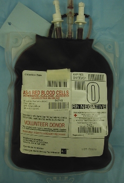
In most other settings, however, whole blood transfusion has largely become a thing of the past. Instead, when blood is donated, it’s immediately reduced to its constituent parts. The red blood cells are pulled out and stored as packed red blood cells (PRBCs); the platelets are pulled out and stored as condensed platelet concentrate; and everything that’s left — the plasma itself, including electrolyte-rich water, clotting factors, immune factors, and other ingredients — is frozen and stored as fresh frozen plasma (FFP). One unit of blood (around a pint) yields one unit of each component. Since most patients only need one or two of these components, we can divvy them out as indicated, and the same blood supply can benefit up to three people.

So for years it’s been standard to transfuse traumatic shock patients red blood cells. As we know, the key problem of shock is inadequate oxygen delivery, and red blood cells are how we deliver oxygen. So drop in a few extra hemoglobin, perhaps top them off with a bit of fluid to keep things moving, and we should be set, right?

Maybe. But this leaves out a number of factors.

First of all, remember our prime directive. Stopping the bleeding is more important than topping off the tanks. How does our body control bleeding? Platelet aggregation and coagulation. And remember that platelets, the bricks of this process, are not reusable; if we have a lot of trauma, and we lose a lot of blood, we can easily run out of them. Does transfusing red blood cells alone provide any platelets? Nope.

So maybe we should throw in some platelets too. But wait — we know that to actually bind the platelets into a cohesive clot, we need a host of backup players, the numerous coagulation factors that live in the plasma. Does a platelet pack provide these? Nope. (Okay, platelets are usually stored in a small amount of plasma, so there’s a few, but not enough.) So maybe we should give the patient some plasma too (or even isolated concentrates of clotting factors to really supercharge the process).

The result of all this is the recent movement towards so-called 1:1:1 therapy, where trauma patients receive equal proportions of red blood cells, plasma, and platelets. In other words, they end up getting all the individual components of whole blood; we just don’t often have whole blood available, or we might give that. This is still an area of active research, and the exact ideal ratios are up for debate; the ratio of red blood cells to plasma is often either 1:1 or very close to it (1:2, 1:3, etc.), and platelets are usually given in somewhat lower quantities, but should not be neglected. The best ratio, as well as the actual quantity of blood to ultimately give, remains to be seen.

Logistics can stand in the way of some of these efforts. For instance, plasma is typically stored frozen (as FFP), and therefore needs to be thawed before use, a process that takes some time. Very large trauma centers may be able to keep a rotating supply of thawed plasma on hand for emergency use, but many facilities won’t be able to have plasma immediately available in this way. And although transfusing in the field seems tempting, the practical challenges of carrying blood products on an ambulance are daunting.

Furthermore, banked blood is not “as good” as the patient’s own blood no matter how it’s given. Even a 1:1:1 transfusion, properly typed, screened, and cross-matched, has real risks of transmitting infection or causing an adverse reaction, carries less oxygen than fresh blood, has reduced hemoglobin pliability (the little disks “stiffen,” becoming less able to squeeze down capillaries to reach the hungry cells), and reduced numbers of labile clotting factors (particularly V and VII). It carries less 2,3-DPG, its pH is lower, and due to the anticoagulants and preservatives added for storage, it’s literally larger and more dilute than the whole blood it started as. Since transfusions are generally not our problem in the field, the applicable moral here is simply that “top ’em up” is not a simple or easy answer to shock, and the only intervention that truly keeps the patient out of trouble is to stop the bleeding!

From the Trauma Professional’s Blog at http://regionstraumapro.com/


In brief:

  1. Blood transfusion is an important step in treating traumatic shock, secondary only to controlling the source of hemorrhage.
  2. Modern “component” blood banking allows for the administration of almost any ratio of red blood cells, plasma, and platelets.
  3. Transfusing primarily red blood cells is the traditional approach, but a movement has recently developed toward more balanced ratios.

Next time: the legacy of crystalloids.

Go to Part VI or back to Part IV

Understanding Shock III: Pathophysiology

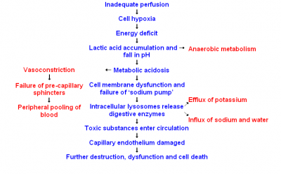
An example of the shock cascade

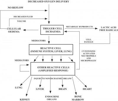
Another model

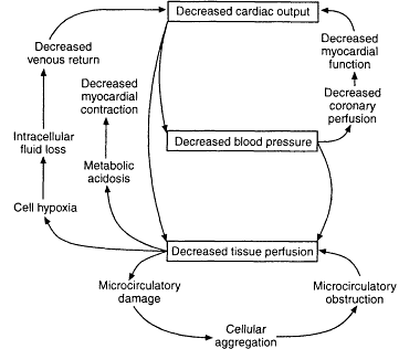
Yet another model


The common thread that defines the shock process is inflammation.

As we know, inflammation is the body’s response to damage. When things go wrong, when trouble calls, we ring the bell for inflammation to make it right. Often this serves us well, but like any militia, if left unchecked it can be worse than the problem it came to fix.

The many twists and turns of the pathology of shock are still not fully understood, but here are some of the important stepping stones along the way:

Shock occurs, and many of the body’s systems are left without adequate oxygen. Although oxygen supplies our primary method of generating energy — the aerobic metabolism — we do have secondary systems in place that can produce energy without oxygen, the anaerobic cycles. In the setting of shock, these take over.

But they’re not great. They provide far less energy than aerobic metabolism, and they produce by-products that accumulate in the body. Among other things, this includes the accumulation of hydrogen ions, creating a widespread acidosis. Think about running sprints or lifting heavy weights; think about that burning feeling, and the eventual failure of your muscles. Operating in an anearobic mode causes trouble and is shortlived at best.

Sooner or later, this isn’t enough to keep things working, and cells begin to accumulate toxic products and eventually shut down. They’re not quite dead yet; they’re hurting, but they can still recover. Like a business that shuts its doors in the off-season, there simply isn’t enough inflow for them to operate right now.

The trouble is, we need those cells. They make up the tissues that form the heart, the brain, the lungs, the kidneys, the liver, and so forth. When the cells close up shop, the organs begin to fail. When organs fail, they cease to provide their essential functions. Let’s consider just one, the heart.

The heart pumps blood. When it loses its effectiveness, it pumps less blood. This means less circulation of oxygen, which means hypoxia is exacerbated. Look at that — we just magnified the problem. If the shock gets worse, is that going to help the heart pump any better? Dream on. The vicious cycle accelerates further.

As hypoxic damage to the cells progresses, the body responds with widespread inflammation to repair it. The trouble is, there’s no real hope of repairing anything without restoring the oxygen supply — but that never stopped Old Man Inflammation. One of his brute-force tactics is to increase capillary permeability, the “tightness” of tissues; everything becomes more susceptible to leakage. The fluid that runs throughout your body begins to ooze everywhere. Generalized edema occurs. In some cases, this is just gross; look at the bloated extremities of the recently dead for an example. But what happens when there’s edema and inflammation of the vital organs? They fail. Fluid in the lungs impairs respiration. Fluid in the brain causes increased intracranial pressure. Another blind response of the inflammatory system is apoptosis, where hypoxic cells — sensing that they’re done for — trigger self-destruct mechanisms and tear themselves apart. Unfortunately, you need those cells.

And hey, what about that acidosis? Our cells (including the ligand-receptor complexes that trigger our sympathetic processes) are designed to function at a specific pH. Placing them in an acidotic environment impairs their function. Combo attack!

But what about our compensatory systems? When our body sees shock, it does things like vasoconstricting, increasing heart rate and contractility, and attempting to maximize the availability of oxygen. That’s great when it works. But when things progress, it’s not so great. Vasoconstriction can choke off the organs, giving them even less oxygenated blood. Tachycardia increases the heart’s demand for oxygen.

And oh, by the way, none of this is adds much to the body’s ability to combat the original cause of the shock, whether that was traumatic injury, a septic infection, or something else.

Key points:

  1. The processes of shock are multiple and self-reinforcing.
  2. Inflammation plays a major role.
  3. Multi-organ dysfunction and failure also plays a major role.

Next time: so what do we do about it?

Go to Part IV or back to Part II

Understanding Shock II: What the What?

. . . the rude unhinging of the machinery of life.

Samuel Gross


When we say shock, what do we mean?

First, to be clear, we’re not talking about “shock” as in “I’m shocked by all this,” or as in “shell shock,” or as in “tasers give an electric shock.” Shock is a formal medical term with a specific meaning.

Here’s the simple definition: shock is what happens when your body runs low on oxygen.

Your entire body, from the top of your horns to the bottom of your hooves, is made of cells. Your cells do various things to keep you alive. In order to do those things, they need a supply of oxygen. Just like your car runs on gasoline or your computer draws electricity, if your cells don’t have oxygen, they don’t work. Essentially, every death, no matter what started the trouble, is caused in the end by insufficient oxygen delivered to the cells.

Without oxygen, eventually your cells die, and then, so do you. However, before that happens, you enter shock.

Mind you that we’re not talking about localized tissue hypoxia. If you tie a tourniquet around your arm, your hand will run out of oxygen and have problems. If a clot blocks an artery in your brain, parts of your noodle will die. These are problems, but they aren’t shock. Shock is a generalized situation; shock happens when hypoxia is widespread and systemic.

Why would such a thing happen? Usually, it happens because there isn’t enough blood flowing to supply oxygen to your organs. Blood is the expressway for oxygen delivery; without enough blood moving at the right speed to all the nooks and crannies of your body, the oxygen won’t get there, and your cells will start to lose their little minds. Blood plays a lot of roles, but this is by far the most important. So although hypoxia is the problem, inadequate perfusion is typically the cause, and we often talk about blood supply as a shorthand for talking about oxygen delivery. There are different types of shock with different underlying causes, but this is the common element that unites them.

Everyone on board so far? If you made it past page 2 of your EMT textbook, you probably knew all of this. But there’s a twist coming, and it’s important. To illustrate it, consider this parable.

You’re shot in the belly, and you bleed out a large portion of your blood onto the ground. We bring you to the hospital, where surgeons repair every inch of damage; you are made as good as new. We replace every drop of blood you’ve lost. At this point, your tissues are repaired, your blood supply is restored, and you’re alive.

But a week later, you die in the ICU.

Why?

The key to understanding shock is this:

Shock is caused by inadequate perfusion, but shock is far more than that.

Say what?

Okay, put another way: no matter what causes the shock, shock leads to more shock.


The shock cascade

When cells become hypoxic, what happens next?

What happens is that they start to do their jobs badly, and this leads to all sorts of systemic problems. When the organs stop working properly, it leads to worsening shock and decreased perfusion, which in turn worsens the original hypoxia, which causes further dysfunction. This process feeds itself.

Dr. Jeff Guy uses this metaphor: suppose you drop a lit match in a dry forest. At this moment, what is the problem? Simple: a burning match. Correcting the problem is equally simple: extinguish it.

But then, the match catches some leaves, and the leaves ignite some dry twigs, and there’s a small fire. What’s the problem? Well, now it’s a little fire going. We can correct it, but we’ll need some blankets or water or well-placed dirt.

What about two minutes from now? The flame has grown, and now it’s a bonfire. We can put it out, but it’ll take some real effort, and it’s going to leave damage.

What about an hour from now? The entire forest is ablaze. The only hope of stopping it will be a massive effort by helicopters and tanker trucks, and even then, most of the trees are probably a lost cause. Maybe we won’t be able to beat a fire that size no matter what we do.

Question: even if we can find that original match in the forest fire, will putting it out extinguish the blaze?

Of course not. The fire has spread.

Shock is a forest fire. The initial hypoperfusion is one thing, and we should try and correct it. But if we don’t, and it starts to cause damage, then that process will start to run away on its own. It will start to cascade, and expand, and feed itself; a new monster is born. Once this has happened, guess what?

We can completely fix the initial hypoperfusion, and still lose the patient.

This happens all the time. Shock occurs, for whatever reason, and we recognize and treat it. But we got there too late. The fire spread. We extinguished the match, but we couldn’t put out the blaze before the damage was too profound to survive. The complications of shock affect nearly every organ system, disrupt nearly every physiological parameter, and undermine the very homeostatic mechanisms that exist to help “fight the fire.” Once this process gets past a certain point, there’s no beating it; the essential fabric of the body is corrupted, and its ability to repair and maintain itself is destroyed. Days or weeks later, despite our best medical care, the patient dies from general, widespread complications. “The operation was successful,” as the surgeons say, “but the patient died.”

That doesn’t mean that we shouldn’t try to fix the initial shock state. That means we should try to fix it immediately.  It means it’s a time-critical, every-second-counts priority — because it’s not the kind of thing we can handle at the last minute. If we don’t nip it in the bud, we’ll go down paths that we can’t come back from.

So, the lessons for today:

  1. Shock is characterized by inadequate oxygen delivery to the cells.
  2. This is typically caused by inadequate bloodflow to the tissues.
  3. Once initiated, shock involves numerous pathological processes that range far beyond the initial hypoxic injury. These complications can persist long after the underlying trigger is corrected.

Next time: a deeper look into some of the “unhingings” that characterize the evolution of shock.

Go to Part III or back to Part I

Differentiating Syncope: A Few Pearls

Syncope. To a fresh-faced student, it’s a snappy word for fainting. To someone with experience, it’s a heavy sigh, because we take a lot of calls for “syncope” and most of them are no big deal. But to a veteran provider, syncope is a deep, dark diagnostic hole—because syncope can be caused by countless different disorders, and although some are benign, a few of them are deadly.

Comprehensive diagnosis and treatment of syncope deserves its own dedicated series, and one of these days we’ll try and work through it from A to Z. Every etiology is unique and has its own distinct pathophysiology, presentation, and treatment considerations. Syncope sucks.

But for now, we’ll just talk about a few take-home pearls that can pay dividends in the everyday management of your next syncope call. We don’t support simplistic rules of thumb ’round these parts, but sometimes 95% of the work can be done by 5% of the know-how, and that’s just fine.

Here are a few dead-simple roadsigns to help guide you through the most common and most important causes of syncope.


Did they pass out and fall, or did they fall and then pass out?

Syncope means that somebody passed out and fell down. It doesn’t mean that they fell down and then lost consciousness. If they tripped on an oil can, fell over and smacked their head on a rock, they may have blacked out, but there’s no mystery there—it’s a simple trauma call.

So, our first step should be to take the raw he passed out and sift it into a more precise description. One problem is that people who lose consciousness often have a poor or unreliable memory of those events, so they may not always be helpful; this is why it’s nice to have witnesses who can tell the story. Of course, witnesses aren’t always reliable either.


Okay, so what do they remember?

To the extent that the patient remembers it, how do they describe the event?

A prodrome is an early, sometimes subtle set of symptoms that warn of a problem developing. Prodromes are our friend, because although they can be very brief or non-obvious, when present they can help indicate what happened. So, ask! It’s the O in OPQRST, and it’s the E in SAMPLE, so it’s the beginning and end of our patient history—no excuses!

Vasovagal syncope is one of the most common causes of syncope, involving a transient drop in blood pressure, and vasovagal syncope is usually preceded by a prodrome. If you’ve never had the experience of standing up too fast and getting briefly faint, here’s the gist: you become light-headed, your vision blurs or darkens, you feel weak, you may stumble, and finally you go down. There may also be broad neurological symptoms, such as visual disturbances (“seeing spots”), strange sensations, shaking, and more. (Basically, your brain isn’t getting enough oxygen, so odd stuff happens.)

How about seizures? Many seizures are preceded by a prodrome known as an “aura,” which can manifest as various unusual neurological abnormalities; read more in our piece on seizures. Did the patient truly lose consciousness, or do they claim that they remained somewhat aware? In a simple partial seizure, the patient will remain aware of their surroundings (although these often don’t cause a “syncopal” collapse); in most others they will experience a gap in consciousness.

Syncope caused by cardiac arrhythmias, such as a run of V-tach or a Stokes-Adams attack, will sometimes be preceded by a palpable sensation of weakness, or palpitations  (“fluttering”) in the chest. However, in many cases there will be no warning whatsoever.


What did the witnesses see?

It’s one thing to hear about a prodrome from the patient, but you may get a different story from the bystanders.

What did they see before he went down? Did he become absent, demonstrate tics or tonic immobility, perhaps complain of an aura? Did he demonstrate obvious clonic jerking of the muscles or urinary incontinence? If he’s acting normally now, was there a period after the event where he demonstrated sluggish activity or unusual behavior, consistent with a post-ictal period? These are all suggestive of a seizure.

Were his eyes open or closed for the duration? Closed is typical of classic syncope, such as a vagal event; open is more appropriate for a seizure. If open, were they rolled back? This also suggests seizure.

Did the patient say, do, or complain of anything before or after the event, which he may no longer recall? Dizziness, headache, chest pain?

Did he stumble, lean against something, or seem to become dizzy? After he went down, did he regain consciousness almost immediately? These are suggestive of vasovagal; once a horizontal position is reached, perfusion to the brain is restored and the problem resolves. If he remained unconscious for a prolonged period while prone—or his initial episode occurred while already seated or reclined—this is highly unusual for vasovagal.

Was he walking and moving normally, in no distress, when he suddenly collapsed like a marionette with its strings cut, hitting the ground with no attempt to protect himself? This is strongly suggestive of a cardiac event and these patients should be considered high-risk for sudden death.


Is there a suggestive history or surrounding circumstances?

Sometimes, the chain of events or the patient’s medical history may suggest an etiology.

Is there a known history of a seizure disorder like epilepsy? How about diabetes? (Take a blood sugar if you’re capable of it; in my book, everybody with an altered mental status is diabetic.) Do they have often pass out or become light-headed?

Have they been eating and drinking as normal? Have they had the flu, and been unable to keep down fluids for the past two days? Were they partying all night? Vomiting? Are they a marathon runner who collapsed in 110 degree weather? Dehydration is a common cause of syncope, particularly in the young, healthy population.

Is there a known condition which may have neurological or metabolic involvement? Cancer with metastases to the brain? A recent infection? A congenital heart condition, such as Long QT, hypertrophic cardiomyopathy, or Brugada? For that matter, are they currently drunk or using drugs? If they take psychotropic or other medications, are they compliant with these, or could there have been an under- or over-dose?

Has there been any recent trauma, such as a fall, motor vehicle collision, or assault with injury?

Have there been repeated lapses in and out of consciousness, rather than a single event? This is an ominous sign suggesting a significant problem.


Are there frank clinical signs that suggest a diagnosis?

This is less likely to be useful than the history, but it can help rule in or rule out major, acute emergencies.

Cardiac abnormalities may manifest with irregular pulses, and active decompensation may be revealed in the blood pressure. Whenever possible these patients should receive ECG monitoring, including a 12-lead. Orthostatic vital signs can be considered if vagal, orthostatic, or hypovolemic etiologies are suggested.

All syncope patients, including suspected seizures, should get a neurological workup, particularly a Cincinatti Stroke Scale.

Respiratory adequacy, including pulse oximetry where available, should be assessed.

Evaluate the abdomen for signs of hemorrhage, and inquire about blood in the stool or emesis as well.