Live from Prospect St: The Big Crunch (conclusion)

Continued from part 1 and part 2


In the end, all three patients receive spinal immobilization. You transport both pediatric patients to Bullitt Medical Center; the P12 assumes care of the mother and transports her to the same destination. No significant injuries are found upon follow-up assessments; however, when the P12 checks Samantha’s blood glucose, they find it to be 32 mg/dL. They administer D50, normalizing her sugar, which improves her level of consciousness; however, she remains confused and becomes somewhat combative. She does endorse substantial alcohol ingestion, is somewhat unclear on drug use, and continues to deny a history of diabetes.

After transferring care, both crews fill out state-mandated documentation to report child abuse, with regard to the mother driving two young children while under the influence and without appropriate car seats or other restraints. You write your documentation with extra caution, aware that it may eventually be used in a court of law.


Discussion

This was a case where no patient was highly acute, but operational issues required some attention and medical confounders obscured the assessment.


General considerations for MVAs

With any significant MVA (or MVC for “motor vehicle collision,” since the DoT takes the position that nothing is truly accidental), there are several factors we should consider:

  • Scene safety. Wherever the scene may be, it’s generally at or near a roadway, and it’s a location that’s already proven itself accident-prone. In this case, we were situated in a truck yard somewhat off the main road. If it were a busier area, and we were first to arrive, we would want to park the ambulance to shield the scene from traffic, and request fire apparatus (for more blocking) and police (for traffic control). We should also consider the presence of chemicals or other hazardous material in an industrial area, which was not a problem here.
  • Extrication. The time to request additional resources is early. Heavy extrication, where vehicle frames need to be bent or cut, is usually performed by fire department ladder trucks or dedicated rescue apparatus; in this case, the driver’s door was dented and needed to be popped open (technically “confinement” rather than “entrapment”), and it was handled prior to our arrival.
  • Cause. Some accidents happen for obvious reasons, such as inattention. Sometimes they’re due to conditions, such as weather or visibility, which is a good clue that such conditions probably persist and might endanger you as well; protect the scene and be cautious during extrication and transport. Sometimes, accidents have a medical cause, which was the case here.
  • Damage. We are clinicians, not mechanics, but vehicle damage can provide clues to injury type and severity. Modern vehicles often develop horrific-looking body damage while yielding minor personal injury; automotive safety science has become quite advanced, and a large part of a car’s protection comes from intentionally crumpling to absorb impact. If occupants are restrained, the vehicle can easily eat up a large amount of shock without anyone suffering significant harm. In this case, we saw a front-left impact at seemingly moderate speed, so we anticipate a head-on type injury pattern with some lateral energy. Damage to the driver’s-side lower dashboard area, plus minor knee injury, suggested a “down and under” rather than “up and over” direction of movement, which is typical for a restrained driver; the windshield was also missing any apparent point-of-impact, which supports this. With the seatbelt and airbag, we were not too suspicious of frontal head injury, but we did look for evidence of lateral head impact against the window or side-wall; we found no obvious head trauma or internal vehicle damage. There was likewise no signs of internal impact from the children in the rear, although we remain suspicious of pelvic or abdominal trauma, since they were wearing lap belts without any torso restraints.
  • Number of patients. Life was made easier by the truck driver, who was obviously unharmed and decided to elope from the scene prior to our arrival. Samantha was making vague reference to her brother, but it seemed that he was coming to meet her and was not an occupant. It is somewhat bad form to forget about people, so it’s good to try and confirm these things, and the first-in responders (the fire department in this case) can help.


Assessment

Just like in most cases, the majority of essential information was communicated in the first few seconds on scene.

Our eyeball exam from twenty feet was enough for an initial assessment on the kids. The Pediatric Assessment Triangle is a model for identifying pediatric life threats that focuses on obvious, big-payoff findings rather than details (like specific vital signs) which can be tough to measure. The three components are:

  • General appearance. This is overall impression and rough neurological status. Are they conscious? If so, sluggish, alert, groggy, engaged with their surroundings, tracking with their eyes? Is there any muscle tone or are they limp? Are they crying? If so, are they consolable? Do they look sick or well?
  • Work of breathing. This is respiratory assessment. Is the child struggling to breathe? Are they tripoding or assuming a sniffing position to maintain an airway? Is there accessory muscle use, pursed-lip breathing, nasal flaring, chest retractions? Are grossly adventitious breath sounds audible (i.e. wheezing, stridor, grunting, snoring)?
  • Circulation. This is general circulatory status. Is skin pink and warm? Is there clear cyanosis, pallor, mottling? Obvious bleeding?

From the first moments on scene, we were able to observe that the pediatric patients were: conscious, crying loudly (therefore with a patent airway and adequate breathing), generally unhappy but not acutely distressed, without obvious bleeding or other trauma, and with normal skin signs. That’s plenty for the initial triage — a more full assessment will come later, but it’s unlikely that we’ll uncover any true life threats.

How about mom? We initially notice no obvious issues except for an altered mental status, which may be masking other problems (such as pain or neurological deficits). We also don’t know the cause of the AMS. Is there alcohol involved? Probably: she directly endorsed this. Drugs? Perhaps: vehemently denying drug use is not uncommon in drug users, and there were purpura consistent with needle “track marks” on her arm. But even if present, neither of those precludes a concomitant traumatic head injury; drunk and high people can bump their head too. And we were reminded of the first rule of EMS: everybody is diabetic. Although the circumstances didn’t necessarily suggest hypoglycemia as the most likely cause, it fit the presentation, and all drunk patients are somewhat at risk for this complication. If she’d stayed in our care, glucometry would have been wise during transport.

Is spinal immobilization needed? Local protocol comes into play. The children are probably low risk. The mechanism as a whole is potentially risky, due to the possibility of side-on energy transfer and head injury, but generally is not too alarming and the assessment findings are fairly reassuring. In the case of the mother, she is the classic example of a poor reporter who cannot reliably describe neck or back pain or participate in a neurological exam; most selective immobilization protocols (such as NEXUS or the Canadian C-spine rule) would advise immobilization in such cases. In this instance, due to equipment shortcomings, one child was immobilized via KED and the other two patients immobilized to long boards, with towel rolls used liberally. The children were liberated almost immediately after arrival at the ED, after a clinical exam by the pediatric emergency physician. The mother began fighting her board after she was roused with D50.


Transport and documentation

This case highlighted the need for intelligent patient assessment to guide transport destinations. Although low-acuity pediatric patients can sometimes be assessed in an adult ED, it depends on the receiving physician’s level of comfort, so in many cases they’ll prefer to transfer them to a specialty center (and any time a patient has to be transferred from where we brought them, we’ve failed them somewhat).

In a similar vein, acute patients needing surgical intervention should always be delivered to trauma centers. Does mom need a trauma center? Since we’re unable to rule out a traumatic cause for her mental status, it’s probably wise, although perhaps not essential. Do the kids need a pediatric trauma center? Probably not; they are, by all appearances, doing fine. Finally, although we could transport parent and kids to different hospitals, it would be distressing to everyone and create logistical headaches (involving consent, billing, and other concerns), so Bullitt Medical Center (an adult trauma center as well as a pediatric ED, although not a pediatric trauma center) is a sensible destination. (Since it’s a larger hospital, it’s also more capable of sustaining the “hit” of receiving three patients simultaneously than a small community ED.) Since the mother is a more challenging patient, it makes sense for the paramedics to take her while our BLS unit acts as a bus for the kids.

As for documentation, depending on state law we may be required to report all instances of child abuse to protective agencies. (In this particular region, reporting is mandated for any child or elder abuse.) If so, local procedures should be followed; although the hospital will most likely perform such reporting as well, in many states this does not absolve EMS of its own responsibilities.

When documenting the call, be aware that charges may be pursued against the mother for neglect, driving under the influence, or other offenses. These may hinge upon your documented findings, such as altered mental status, lack of appropriate child restraints, or statements about substance use. Depending on local laws for mandated reporters, you may be required to report these findings directly to police, or you may actually be prohibited from doing so by HIPAA laws; in either case, however, they should be noted in your report.
Live from Prospect St: The Big Crunch (part 2)

Continued from Part 1

Since the two children appear generally intact, you ask your partner to evaluate them more fully while you head for the sedan to find the driver. Anticipating three transports, two stable and one potentially critical, you ask your dispatch to continue the P12, and also to ensure that police are en route (they are).

Arriving at the sedan, you find a middle-aged woman in the driver’s seat, alert. She is pink and warm, perhaps more diaphoretic than you’d expect for the ambient temperature, and does not initially notice as you kneel beside her. A firefighter is holding C-spine immobilization from the back seat.

When you greet her and pat her on the shoulder, she gives no response, but with more vigorous stimulation she looks over and acknowledges you distractedly. With multiple attempts and some yelling, you’re able to get answers to a few questions, but she is slow, tangential, and often ignores you outright. She gives her name as Samantha, but cannot or will not provide her last name; she is unable to describe the events that led to the collision; and she gives no medical history or current medications. She does state several times that she’s fine and would like to leave. When asked about her passengers, she mumbles “my kids” and mentions her brother several times. She endorses pain when asked explicitly, but does not specify where. She agrees that she drank “a little” alcohol; when asked about any drug use, she denies it vehemently.

Physically, she appears generally unremarkable. She is breathing somewhat shallowly but effectively, and her radial pulse is around 100 and slightly weak. Her seatbelt is not in place, but it’s unclear whether it was removed at some point. No gross trauma is apparent upon her head, face, or neck, and she does not complain or grimace upon palpation. She is uncooperative with a neurological exam, but demonstrates spontaneous movement of all four extremities. Her pupils are equal and seem appropriately small on this moderately bright day. Chest rise is generally equal and her abdomen is supple; no bruising consistent with seatbelt injury is visible. Her left knee is abraded and somewhat swollen. A sprinkling of dark blotches and streaks are noted on her left ventral arm in the antecubital region. Both frontal airbags are deployed; the windshield is cracked, but lacks a “starred” point of impact; and the plastic dashboard in the driver’s knee area is damaged and cracked. No blood or other damage is visible in the interior compartment. There are no child seats.

Your partner comes over. “The kids seem fine, just upset. One’s complaining of some abdominal pain, but it looks okay. They’re little troopers. Fire says they were wearing regular lap belts with the shoulder strap tucked behind them.”

When you wonder aloud whether there are more patients, he says, “There was nobody else in the car when fire arrived. The truck driver gave a statement to the police about how she was swerving across the road and plowed into him, but then he eloped.” He looks over your shoulder. “Oh, and the P12 is pulling up now.”


What is your treatment plan for these three patients? What are their respective priorities, any points of concern, and how could you shed additional light on their status?

Who will transport which patient, and to which destinations?

What special considerations should be made during documentation?


The conclusion is here

Live from Prospect St: The Big Crunch (part 1)

It’s 4:00 PM on a gloomy Friday in Chandlerville, and you’re the technician for the A2, a dual-EMT, transporting BLS unit dedicated to the city. Chandlerville is a small town, but densely populated, and its numerous industrial districts are frequent sources of work. 911 dispatch is directly through the fire department, which also sends a BLS fire apparatus to assist on all medical calls; your company’s ALS is also available by request. You are equipped with finger-stick glucometry, glucose, aspirin, and epinephrine.

After a “man down” call that ended in a patient refusal, you’re now returning to quarters. Just as you’re beginning to back into the garage, a tone sounds.

Engine 3 and Ambulance 2, respond to 2108 Coastal Rd, the Empire Shipping Company, for an MVA. That’s two-one-oh-eight Coastal Road, in front of Empire Shipping, for an MVA. Engine 3?

“Engine 3 is responding.”

Ambulance 2?

As your partner flips on the lights and pulls out to the street, he speaks into the radio: “Ambulance 2 has 2108 Coastal Rd.”

Time out 16:01.

Coastal Road is a long connector that wraps around the edge of town, and you glance at the map book to confirm that the 2000 block will be near the very end, about as far away as you can get in Chandlerville. Engine 3 is stationed in that district, however, so they arrive within minutes.

“Engine 3 to Firecom.”

Firecom answering.

“We’re off at 2108 Coastal Road. Two-car MVA, car versus truck. Multiple injured parties and entrapment. Start an ALS unit and a ladder for extrication.”

Engine 3, you have a car versus truck, multiple injuries with entrapment. Break. Ladder 3, respond to 2108 Coastal Rd for the MVA; Engine 3 is on scene and A2 is responding. Time out 16:04.

A few seconds later, your company radio dispatches Paramedic 12 to the same address, after Chandlerville Firecom contacts them via landline. The P12 starts responding, but they’re coming from two towns away, with an ETA of 10+ minutes. The field supervisor also starts rolling from an unknown location to assist. 30 seconds later, Engine 3 updates that they have an injured adult and several children.

Now very awake, you reflect that the nearest hospital will be Chandlerville Memorial, a 3–5 minute emergent transport (10 minutes otherwise). The nearest large tertiary center, Bullitt Medical Center — a Level I adult trauma center and a designated pediatric ED — is 15 minutes emergently (25 otherwise). The nearest Level I pediatric trauma center, however, is the Children’s Hospital, which is also 15 minutes but in the opposite direction; they do not receive adult patients.

Ladder 3 arrives on scene momentarily, and you pull up a few minutes later. As you park and call yourself out, you observe a Ford sedan with its front left corner smashed in, two feet of its fender and frame crumpled. This is evidently the result of driving almost headlong into the side of an 18-wheeler. It appears that the driver swerved right to avoid the truck, undercutting its rear wheels and “submarining” itself; the damage reaches the passenger compartment, but there does not appear to be significant intrusion. The truck itself seems minimally damaged.

As you jump out, a firefighter waves you down. “We’ve got three!” he announces. “Mom’s in the driver’s seat; she seems really loopy, probably drunk. Her door is just dented, we popped it open. But her kids are over there.”

Twenty feet away, you see two young girls, around 4 years old, each in the arms of a firefighter. They are crying loudly and clearly upset, with no visible injuries. The mother is hidden from sight in the sedan. The driver of the truck is nowhere to be seen.


What are your initial steps for addressing this scene?

Who appears to be the first priority for care?

What resources will you need? Which, if any, should you cancel?


Continued in part 2 and the conclusion

Glucometry: Introduction


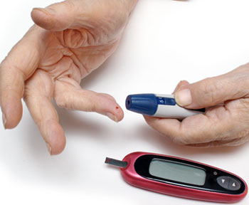
Glucometry — i.e. bedside measurement of blood glucose levels, usually from a capillary finger-stick — is an ALS skill almost everywhere, and in some systems it’s available to BLS providers as well. Even in places where it isn’t technically permitted for BLS, it’s often still widely allowed on a “wink and nod” basis, especially on mixed-staffing units where the paramedic has better things to do than apply droplets to test strips.

In other words, it’s something we do. Moreover, it’s something very valuable that we do. I work in a system that allows BLS glucometry along with various other “extras,” and if I had to give up all of it (nebulized albuterol, nasal naloxone, and more) to keep the glucometer, I’d do it in a heartbeat. It serves an invaluable and often irreplaceable role in patient assessment, and it’s used often, not sometimes.

As with any tool, though (such as pulse oximetry), intelligently using the device requires understanding how it works, how its results should be clinically applied, and when it fails. Unfortunately, this is rarely taught in depth, beyond perhaps a brief “How’s how you press the buttons” in-service. So let’s talk about glucometry. And talking about glucometry means starting with glucose.


Glucose Physiology

Practically speaking, glucose is the fuel of human life.

What’s a fuel? Imagine I’m starting a campfire. I build a pile of wood, I light it with a match, and it begins to merrily burn. As any firefighter (or Boy Scout) knows, a fire needs certain things. It won’t burn without a supply of oxygen. It won’t light without a heat source. And, of course, it needs something to actually burn — a fuel.

Although humans have a few different metabolic processes that allow us to survive in difficult circumstances, for the most part, we work the same way as the campfire, except our fuel isn’t wood: it’s glucose. We make a pile of glucose, mix it with oxygen, “light” it with some excess energy, and we’re rewarded with an outpouring of energy far greater than we put in. It’s called aerobic respiration, and almost all of the energy we need to live (sing, dance, hunt the mammoth, think about cellular metabolism, buy cheeseburgers) is generated in this way.

Glucose comes from food; in other words, we eat it. Glucose itself is a very simple sugar, and generally, we don’t literally spoon glucose into our mouths; instead, we eat more complex foods (like cheeseburgers), and our bodies break them down or transform them — either directly into glucose for immediate use, or into a form we can store (like fat), which can be readily broken down to burn later.

Remember, folks: the basic “fire” of life burns glucose and oxygen. We know that without oxygen, we quickly die. For the exact same reason, without glucose — we die. This is not optional stuff, and the only reason we survive longer without cheeseburgers than without air is because our bodies can store substantial amounts of fuel for later use, whereas we can only retain a few minute’s worth of oxygen. (We can also generate some energy through anaerobic metabolism, an “oxygenless fire,” but only very little; it’s a short-term reserve that burns out fast.) Every cell in the body therefore needs a constant supply of fuel to keep its machinery running, and this is supplied by glucose circulating in the bloodstream. Since this stuff is so important, our bodies are very good at monitoring the amount of circulating glucose, replenishing it from reserves when it’s low, and dumping it off when it’s high.

One of the main ways that this fine-tuning is done is using a hormone called insulin. Glucose needs to enter cells in order to be burned, but we want tight control on how much of it enters at any given time (since we need to keep enough circulating for the rest of the cells), so access is managed by a “lock-and-key” mechanism. To pass into most cells, glucose needs to “unlock the door” using an insulin “key.” Without insulin, we can have all the glucose in the world circulating through the blood, but it won’t be able to enter the cells hungry for it, any more than you can get into your house to feed your cat if you’ve lost your keys.


Diabetes

Now, let’s say that I have glucose in my blood. And I’m releasing insulin to let it access my cells. But my cells aren’t listening. It’s like somebody changed all the locks on me; we still have the key, but suddenly, it’s no longer opening the doors.

Why would this happen? There are various reasons, including genetics, certain medications, and a few diseases. But often, a key factor is habituation. If we keep our glucose levels elevated all the time (say, by eating a lot of rapidly-digested sugars), then our insulin levels will also be elevated all the time, and eventually, the insulin receptors on the cell membranes will say: “Boy, it seems like there’s a ton of this stuff around; I must be too sensitive to it. I’ll start ignoring some of it.” This is called insulin resistance, and it can range from mild (only some receptors of some cells are a little resistant) to severe (most cells are practically ignoring insulin). Unfortunately, this problem tends to exacerbate itself, because when our control centers see that releasing insulin isn’t lowering the circulating blood glucose as much as it should, we release more insulin, which encourages further insulin resistance… and so on.

The result of this is that more glucose tends to remain in our blood than we need: hyperglycemia. This isn’t a good thing; all that extra sugar zooming through our veins has a habit of piling up in the wrong places, which leads to strokes, heart attacks, pulmonary embolisms, DVTs, peripheral vascular disease, kidney failure, and more. It sucks, and it’s called type II diabetes mellitus. (Mellitus refers to sugar, and it distinguishes DM from diabetes insipidus, a totally unrelated disease.)

On the other hand, what if we can’t make insulin at all? Usually, this happens when our body’s immune system attacks the emitters that produce insulin, for unclear but unfortunate reasons. It usually begins when we’re young, and although it can be precipitated by various triggers, it generally happens more or less on its own. Whatever the case, if we can’t produce insulin, we’ve lost the key, and glucose can’t enter our cells. Without glucose, the fire doesn’t burn, and we die. It’s called type I diabetes mellitus, and without treatment, it’s always fatal.

Nowadays, type I diabetics survive by taking exogenous insulin — since they can’t make their own, we synthesize it for them, and they simply inject it. (They still make their own insulin, so most type II diabetics don’t need to inject the stuff; they manage their blood sugar through careful control of how much they eat. In some cases, however, particularly in the elderly or anyone who is less able to tightly manage their diet, type IIs will also use insulin to help adjust their levels.) How do they know how much to take?

There are ways to estimate insulin doses by, for instance, measuring how much food you’re eating, or from past experience. However, it’s also incredibly easy to misdose. Insulin is a powerful, powerful drug, and a small change in dose can mean the difference between bringing you to a normal, healthy blood sugar, and sucking every last glucose molecule out of your blood until you’re dangerously low — hypoglycemic.

Although hyperglycemia is unhealthy in the long run, and massive hyperglycemia can be an acute danger, even brief periods of modest hypoglycemia can be deadly, so it’s something to avoid. As a rule, the problem in diabetes is too much sugar, not too little, so left on their own, almost no diabetic would become hypoglycemic. However, since all type I and some type II diabetics take exogenous insulin, hypoglycemia happens all the time due to overdosing. In other words, we do it to ourselves — or to our patients — accidentally. (Even when type IIs don’t take insulin, they almost always take other drugs that help mitigate glucose levels or sensitize their insulin receptors, and some of these meds can also cause hypoglycemia.) Getting it right isn’t as easy as it sounds, because numerous factors can cause changes in your blood glucose and/or your insulin sensitivity; for instance, exercise depletes glucose (the hotter fire needs that fuel), so if you hit the gym and forget to eat more or to reduce your dose to compensate, you can easily deplete your available sugar and collapse.

The best way to get the right insulin dose is to accurately track your current blood sugar, and nowadays, this is done easily and quickly using a hand-held glucometer. Tune in next time, and we’ll talk about how they work and how to use them.

Continued in Glucometry: How to Do It and Glucometry: Clinical Interpretation

What it Looks Like: Cardiac Arrest and CPR

Update: Our friends at EMS 12 Lead have put together a “sister post” to this one, with further discussion and some additional clips. Check it out!


Although we’ve talked about the fundamentals of good CPR before (and then again), the fact remains that the first step of any resuscitation is recognizing the presence of cardiac arrest. In fact, failure to do this in a timely fashion is a common problem at all levels of healthcare: because these situations don’t happen often, we are reluctant to accept when they’re happening now. (Real emergencies don’t come heralded by a change in soundtrack.) The result is delays, often for many minutes, before anybody initiates CPR and attempts defibrillation. We can’t just point fingers at the bystanders and lay providers — it’s also the EMTs, the nurses, even the doctors doing this. “Is that a pulse?” we muse. “I think there’s a pulse. Here, come feel.”

It’s true that cardiac arrest, at least in the early stages, is often not easily distinguished from other maladies (such as unconsciousness due to seizure or drugs). A few clues may be immediately obvious, such as pallor of the skin if some time has passed, or if a bystander actually witnesses the patient suddenly collapse. However, in the end, the way to make this call quickly and reliably is to simply follow the algorithm. You’re not the first person to deal with this, and the American Heart Association has spent years simplifying the decision process — because the goal isn’t to eventually “figure it out,” the idea is to immediately recognize it and start lifesaving measures within seconds.

Is the patient responsive? (No; they appear unconscious, and make no response whatsoever to painful stimuli.) Are they breathing normally? (No; they’re not breathing, or merely performing agonal, “gasping” breaths.) Is there a carotid pulse? (No, no pulse is palpable within a few seconds.) That’s good enough for us. Start pushing on their chest and don’t stop unless it’s absolutely essential — and the only things that are absolutely essential are checking their cardiac rhythm (just a few seconds) and delivering a shock (less than a second).

We’re going to look at a number of examples of real-life cardiac arrest (or “codes” in the usual lingo). As a rule, the actual CPR that you’ll see here is of relatively poor quality. This is due to a number of factors, but primarily it’s because 1) Many of these clips are five, ten, or fifteen years old, from a time when CPR was taught and practiced differently; and 2) Even today, many people do not perform good CPR.

So: focus on the patients. Watch how they present, their breathing, their skin, their responses to the interventions. Watch the challenges that the providers face as far as managing the patient and the environment. Watch how their approaches differ by region, circumstance, or personal preference. But for the most part, do not do what they are doing. We’ll watch a couple examples of really good CPR at the end so you know what to strive for.


We’ve linked this before, and for good reason; it’s one of the best videos I know of a real code. This is older CPR, with less emphasis on compressions and more on ventilation, but otherwise fairly true to the textbook. Notice the early “activation” of EMS, and the brief pulse check. Notice how rather than trying to “one-man” the BVM, they take advantage of the many available hands, allowing one person to hold the mask and one to squeeze the bag. Notice how they quickly dry the chest for the AED without being obsessive about it. As for the compressions, nowadays we would like to see them faster and deeper, with fewer and briefer pauses.

In the patient, watch the spastic, gulping movements of the mouth and tongue; this is agonal breathing. Notice also the decorticate posturing of the upper body, suggesting neurological dysfunction. Finally, notice how (after the third round of CPR + defibrillation), he begins to breathe spontaneously, with obvious chest rise, and this is clearly different from the prior agonal respirations.


(watch through 8:45) Despite the numerous pauses for commentary, this is also good. The initial compressions are rapid — a little too rapid, which is okay, but not deep enough, and if they were deeper they would likely be at a more reasonable rate. The second compressor goes deeper, but does not recoil fully at the top. The third (male) rescuer gives perhaps the best compressions, but notice his elbows — although pushing hard and deep, he allows his elbows to bend slightly each time. This is a very common error in otherwise skilled compressors, and is a good way to fatigue yourself quickly. Make a conscious effort to lock the elbows out completely, allowing you throw your full weight behind each compression rather than “pressing” with the arms. Notice also how frequently the rescuers stop compressions for one reason or another. Chest compressions need to build upon each other for several compressions before you’re producing anything like the coronary perfusion pressures you want to see; repeatedly stopping and starting sacrifices all your hard work.

In the patient, notice the pallor (paleness) of his skin, and the total lack of tone (limp flaccidity) of his body. Notice how he convulses with the shock, and how his chest rises and expands with ventilations. Finally, notice how his abdomen recoils outward in a seesaw manner with each downward compression of the chest.


(watch through 7:10) This is a chest pain patient that codes on camera. Despite the low image quality, notice how poorly he immediately presents; he is obviously fatigued, wan, and struggling with some sort of pain or other internal distress. Upon attempting to stand, he loses consciousness and demonstrates agonal respirations (listen to the heavy snoring). They ask if he has a history of seizures; a substantial number of cardiac arrests are initially mistaken for seizures, and may present with seizure-like activity (such as foaming of the mouth). There is obvious difficulty with compressions due to the high position of the stretcher. Bubba was very fortunate to arrest in the immediate presence of paramedics.


(watch through 3:43) Notice again the initial hesitation due to bystanders believing a seizure is occurring. These compressions have the kind of violent depth we want, although at about half the rate. Notice again the slight arm bend.


A chest pain patient who deteriorates into a full arrest while on camera for a UK documentary. Depicts a good portion of the code.


[Added 5/8/13 — ed.]

(watch until the credits)

ED footage of EMS bringing in a code. Shows the practice of “code surfing,” where a rescuer rides the stretcher to provide ongoing compressions during movement — a great idea if you can do it safely and effectively (it helps to use someone small!) Notice how fast some of the compressions are performed, but it’s tough to reach good depth at those rates, particularly when the arms aren’t held straight. Although the captions note that the patient had ROSC, it’s extremely unlikely that he survived to discharge; when patients are transported without achieving ROSC in the field, they almost never walk out of the hospital. Cardiac arrests are worked on scene; transport without a pulse is simply giving up, unless you have good reason to think there’s a reversible etiology of arrest that the hospital can address.


[Added 8/21/12 — ed.]

(watch through 12:05, or stay for some bystander interviews) Another near-drowning. Decent-looking compressions and a reasonable attempt to minimize interruptions, although notice the pauses for intubation and at various other times. Unknown outcome.


(watch through 2:25) This is a volunteer crew from AMR’s disaster response team in Haiti. There seems to be initial confusion about whether the patient is pulseless or merely apneic, hence the initial focus is on the airway; nowadays we would frown upon interrupting compressions for intubation, and the bagging after the tube has been placed is far too fast (every 6-8 seconds only, please). The teamwork is good, and return of spontaneous circulation (ROSC) is achieved after a few minutes. Notice the decision to defer a blood pressure measurement, since the patient has a strong radial pulse — an indicator of a decent pressure, if not an exact number. The patient does have fixed and dilated pupils, indicating a probable poor neurological status.

Keep watching only if desired; the patient is transported to the field hospital, where she rearrests, and the doctor there halts resuscitation efforts.


(watch through 23:50) This is a neonatal resuscitation immediately following a field delivery of twins; one infant is apneic following birth. BVM ventilations and compressions are performed, as well as an aborted attempt at intubation; however, in the end the neonatal fundamentals of warming, suctioning, stimulation, and supplemental oxygen end up effectively reviving the child.


http://www.youtube.com/watch?v=afo3-dhRnA0

[will not embed; click through to view video then return] Another infant resuscitation, this one in the ED. Excellent footage of compressions, ventilation, and the typical hubbub of a code, as well as an IO (intraosseous) line that infiltrates and the use of ultrasound to assess for cardiac function during PEA.


CPR on a near-drowning. A fine example of the typical poor quality of bystander compressions; notice the negligible depth and general uncertainty about whether to intervene.


A collapse at a sporting event. There is no backstory available on this, so it may not be a true arrest, but if so it would be consistent with commotio cordis, when a blow to the chest (such as a punch) causes an arrhythmia (due to an R-on-T induced by the physical blow; this is the evil brother of a precordial thump, with the opposite effect). This type of arrest has extremely good prognosis for recovery if immediate CPR and defibrillation is performed, since there may be little to no underlying disease; it’s a healthy young patient who simply got whacked wrong.


(watch through :38) Some brief miscellaneous footage of an arrest post-drowning, with a few pretty good compressions.


(watch through :57) Another near-drowning. Nice compressions. Notice the pallor and lack of tone.


[Added 10/11/13 — ed.]

This is clearly an old video, although it’s not clear from what year. Regardless, it’s a great opportunity to list the things you’d do differently today. Since we know that the keys to a successful resuscitation are immediate, deep, fast, uninterrupted compressions, along with rapid defibrillation, do you think this patient had a good outcome? How many of the interventions they performed instead of that stuff are still recommended care? If you were on that scene, would you be an advocate (some might say a CPR Nazi) to ensure that things were done properly?


Finally, let’s look at a couple examples of really spot-on, perfect resuscitation. Since perfection is rare in life, and having a camera in the room is even rarer, these will be simulations.

Click here for a teaching video from the Austin/Travis County medical director’s office. It demonstrates their “pit crew” model, where each member has a designated role, and each action is carefully crafted to match the latest evidence for best practices to promote survival. Notice how compressions begin almost immediately, once the rescuers have noted a lack of responsiveness, breathing, and pulse — and compressions stop for almost nothing, no matter what else is happening. (I would call these compressions very good, but a bit fast and shallow.) Secondary tasks like bagging can happen in the background. This crew does stop compressions while the AED charges, while I personally prefer to compress during this interval (between analysis and shock); the longer you delay between last compression and delivery of the shock, the less chance of getting a pulse back.


(Watch from 2:45 onward) This is the model from Salt Lake City Fire, portraying a highly progressive model. Aside from the general concepts of “compression-centered” resuscitation and the pit crew model, they’re also eliminating pauses for rhythm analysis (using the “see-through” filter on the Zoll monitors, which removes CPR artifact) and even for defibrillation (shocking without taking hands off the chest, which has not been proven safe, but generally seems to be). In other words, there’s essentially no interruption in compressions until there’s evidence of a perfusing rhythm. Notice the compression technique, where knuckles remain against the chest to lock-in the hand position, but the heel of the palm comes off at the top, ensuring full recoil. Beautiful stuff.


There you have it, folks: what dead people look like, and what it looks like when we try to bring them back. Typically the process is chaotic, and we do our best, but often drop the ball on what’s important. Nobody’s perfect, but we can direct our focus toward the pieces that matter the most, and this lets us “streamline” our efforts away from the distractions and toward the critical elements. Recognize the problem early, compress hard, deep, and fast, and don’t stop for anything unless it’s defibrillation. Ain’t so hard, is it?


Sincere thanks to James Oz (Melclin) for assistance with compiling these video clips.


Check out also what Jugular Venous DistentionSeizures, and Agonal Respirations look like