CPR for Dummies: How to Save a Life

One of the peculiarities of EMS education — and as a byproduct, of EMS practice and culture — is that we spend the majority of our time focusing on the minority of our calls. Think about it: your textbook has pages and pages devoted to ruptured aortic aneurysms, placentas previa, and mid-femur fractures — and when’s the last time you saw one of those? But scarcely a paragraph is given to the routine transfer, the drunk asleep on the sidewalk, or the MVC with minimal injuries. Call it an inverted pyramid: the most important stuff is low-volume, the most common stuff is pretty easy.

Whatever. The point is, at the very apex of this pyramid is the cardiac arrest. In its purest form, cardiac arrest is exactly why EMS exists. It couldn’t be higher stakes — as a disease, it’s absolutely certain to be life-threatening — and it’s terribly time sensitive, but the potential exists for a total cure if everything goes well.

Unfortunately, like many low-probability calls, we don’t get a great deal of experience with these — even less if your shift isn’t dedicated to emergencies. And when we don’t get much experience with something, that’s when training needs to fill in the gaps.

CPR and BLS resuscitation can seem like a confusing topic, especially given the frequent and seemingly arbitrary changes to the guidelines. The truth is, though, that it’s only gotten simpler and simpler — and you don’t need to follow the research (read: be a giant nerd like me) in order to know exactly what to do. Here’s the short, stripped-down, painless rules for how to save a life.


Push and Zap

Basically, after around sixty years of research on resuscitation, there are only two things that we know for sure help people survive cardiac arrest: chest compressions and defibrillation.

Literally, just those two things. Oh, there’s other stuff — ventilation, drugs, devices — that seem to help briefly, but so far nothing else has been proven to get someone’s heart beating again and let them walk out of the hospital with a working brain. Now, some of those other things do seem like pretty good ideas, and in many cases we started doing them before we knew if they’d really help or not, so we’re still doing them because people are used to it; it’s part of our training, and it’ll take some extra-compelling evidence to make us actually stop doing that stuff. But still, the story so far: chest compressions and defibrillation definitely help people survive, and that’s it.

What this means is that they should be your number one priority. If your patient is in cardiac arrest, that’s what they need. Other stuff? It may or may not be helpful; if you have the chance, or the personnel, and it doesn’t interfere with chest compressions and defibrillation, then you could go ahead and do it. It might help. But delaying or stopping the big two for that other stuff is like making a thirsty man wait for a drink of water while you comb his hair.


Early, Hard, Fast, Uninterrupted, and Full Recoil

Okay, so, chest compressions. Easy enough. Anyone can do ’em, all you need is your hands, just jump in there and push.

However, that’s not quite the whole story: the quality of compressions matters a great deal. We are literally pumping blood here; we are creating mechanical pressure to replace the squeezing of the heart. Just like you can wriggle a bicycle pump ineffectually without making much progress on inflating your tires, so too can you make goofy movements on someone’s chest without providing much perfusion. Even at its best, CPR only provides weak circulation compared to a real heartbeat; if you give poor CPR that’s even worse.

So here are the key components:

  • Early: Compressions should be initiated as soon as possible after arrest. That means, if I go down now, ideally you’ll start pushing on my chest as soon as I hit the ground. Typically that’s not possible, but mere seconds really do matter here; the longer there’s no circulation, the more tissue is endangered (all tissue, but particularly the vulnerable heart and brain), and the less likely that defibrillation will be successful — or if it is, the more likely there will be permanent complications.
  • Hard: Good chest compressions are a violent, aggressive act. We now recommend a depth of at least 2 inches in adults, which if you examine a mannequin (or fellow human) is remarkably deep. (Yes, “at least” means that going deeper is fine; compressions that are “too deep” are rarely seen in real life.) This isn’t a gentle cardiac massage, it’s not the mellow bouncing you usually see in movies, it’s a deep, powerful, oscillating thrust. It should tire you out, which is why we recommend changing personnel frequently; even when you think you’re still doing well after a few minutes, you’re probably not.
  • Fast: The recommended rate is now “at least” 100 compressions per minute. Since nobody knows what this means without a metronome, I highly recommend “musical pacing,” or using the beat of a well-known song to learn the rate. Stayin’ Alive by the Bee Gees is the classic; I like Queen’s Another One Bites the Dust myself. Again, 100 is an “at least” rate, so faster is better than slower. Admittedly, if you go extremely fast the heart won’t have time to fill between squeezes, but most “ludicrous speed!” CPR tends to have poor depth, and self-regulates anyway once you get tired.
  • Uninterrupted: Just like it’s essential to begin compressions as soon as possible, it’s equally essential to stop them for nothing. It’s not just that every moment you spend off the chest is “dead time” in which no blood is circulating; it’s worse than that. Chest compressions need to generate some “momentum” in order to create enough pressure to perfuse the heart; several consecutive compressions are needed before you’re really moving much blood at all. If you keep stopping — and studies show that everyone stops far more than they realize, to fiddle with one thing or another — you’re wasting those gains as soon as you’ve achieved them. Maximizing this “compression fraction” should be a primary goal; once you get on that chest, don’t stop for anything else unless it’s literally more important than circulating blood.
  • Full recoil: Among otherwise skilled rescuers, one of the most common errors is failing to allow for full recoil of the chest. In other words, you press down deeply, but rather than releasing fully, you start the next compression before you’ve come all the way up. This shortens the stroke of the pump just as much as if you were giving shallow compressions, and for several complex reasons (in particular the loss of preload) can reduce circulation in other ways too. We do this one particularly when we start to get tired, and begin to leaaaan forward to rest on the chest.

Defibrillation

It’s really as simple as this: once the heart’s entered fibrillation (or to a lesser extent a pulseless V-tach), the only plausible way to fix it is with electricity. These people are not going to “come to”; they are not going to have a Baywatch moment where they cough out water and wake up, even if you give them great CPR. They have an intractable problem, and the cure for it is an electric shock. Defibrillation is life-saving.

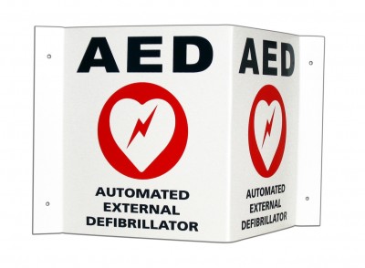
For most of us, this means using an AED, the automated devices you see everywhere from airports to ambulances. The reason they’re everywhere is because their use is time-sensitive, and if you drop dead ten miles from the nearest one, it might as well be ten light-years. No matter where you are, compressions must be performed to buy you time, and a defibrillator must be found to shock you back. If both don’t happen quickly, you will probably stay dead forever.

There are argument about some of the technical aspects of defibrillation, such as pad placement and waveform, but so far none of these details have proven to be very important. What is important is that you shock early, and get ready to shock without interfering with those compressions. Whenever possible, while one person gives compressions, someone else should clear off the chest by cutting or pulling the shirt from under the compressor’s hands, place the pads around them, and start the AED’s cycle. For many models of AED, there will be a period of several seconds while it walks you through voice prompts (telling you to stay calm, call for help, etc; these devices are designed to be usable by laypersons with no training), which should be ignored while you continue your CPR.

Once the AED tells that it’s analyzing the rhythm, you will need to stop compressions; this is the computer’s opportunity to decide whether the patient can be shocked or not, and interfering with this will just delay the process. If it doesn’t advise a shock, get back on the chest; you may have better luck later. If it does advise a shock, get back on the chest anyway! It’ll need to charge first, which may take quite a few seconds, and remember — every second matters. (Just make sure the whole team’s on the same page here, so that nobody pushes “Shock” until you’re clear.)

As soon as the AED announces that it’s ready to shock, everyone should be ready: cleared from the patient and prepared to shock. In a coordinated fashion, the compressor should clear the chest, the shock should be delivered, and he should immediately resume compressions with a pause of only a second or two. Rinse, lather, repeat.

When do you stop this process? When someone much smarter than you says to stop; or when the patient demonstrates clear signs of life (such as movement, breathing, or improved skin signs — or for the medics, a spike in end-tidal CO2). Don’t keep stopping to palpate pulses and otherwise fiddle with the patient. Like a soufflé or a Schroedinger’s cat, you must have faith in the process here, because checking on the process will assuredly cause it to fail.


It Ain’t Rocket Science

People, there are other details to this process, which is why they make us take CPR classes and carry the little cards around. And in 2015, there might be some new ideas on how we can do it best. Research continues apace in the countless EMS systems around the world that are experimenting with different technologies, techniques, and methods to improve survival. That’s how we’ve come from 1–2% survival rates to the 50%+ that a few cities now enjoy. It’s slow going, but it’s going.

But the best methods won’t matter if you don’t use them, and a lot of effort has been given to make our current methods truly simple. You literally can’t go wrong if you give great compressions and defibrillate as soon as possible. You can certainly go wrong if you forget that those are the two most important, life-saving measures — but you’d never forget that, would you?

Push and zap, folks. It’s so easy, an EMT can do it.
Differentiating Syncope: A Few Pearls

Syncope. To a fresh-faced student, it’s a snappy word for fainting. To someone with experience, it’s a heavy sigh, because we take a lot of calls for “syncope” and most of them are no big deal. But to a veteran provider, syncope is a deep, dark diagnostic hole—because syncope can be caused by countless different disorders, and although some are benign, a few of them are deadly.

Comprehensive diagnosis and treatment of syncope deserves its own dedicated series, and one of these days we’ll try and work through it from A to Z. Every etiology is unique and has its own distinct pathophysiology, presentation, and treatment considerations. Syncope sucks.

But for now, we’ll just talk about a few take-home pearls that can pay dividends in the everyday management of your next syncope call. We don’t support simplistic rules of thumb ’round these parts, but sometimes 95% of the work can be done by 5% of the know-how, and that’s just fine.

Here are a few dead-simple roadsigns to help guide you through the most common and most important causes of syncope.


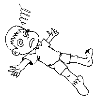
Did they pass out and fall, or did they fall and then pass out?

Syncope means that somebody passed out and fell down. It doesn’t mean that they fell down and then lost consciousness. If they tripped on an oil can, fell over and smacked their head on a rock, they may have blacked out, but there’s no mystery there—it’s a simple trauma call.

So, our first step should be to take the raw he passed out and sift it into a more precise description. One problem is that people who lose consciousness often have a poor or unreliable memory of those events, so they may not always be helpful; this is why it’s nice to have witnesses who can tell the story. Of course, witnesses aren’t always reliable either.


Okay, so what do they remember?

To the extent that the patient remembers it, how do they describe the event?

A prodrome is an early, sometimes subtle set of symptoms that warn of a problem developing. Prodromes are our friend, because although they can be very brief or non-obvious, when present they can help indicate what happened. So, ask! It’s the O in OPQRST, and it’s the E in SAMPLE, so it’s the beginning and end of our patient history—no excuses!

Vasovagal syncope is one of the most common causes of syncope, involving a transient drop in blood pressure, and vasovagal syncope is usually preceded by a prodrome. If you’ve never had the experience of standing up too fast and getting briefly faint, here’s the gist: you become light-headed, your vision blurs or darkens, you feel weak, you may stumble, and finally you go down. There may also be broad neurological symptoms, such as visual disturbances (“seeing spots”), strange sensations, shaking, and more. (Basically, your brain isn’t getting enough oxygen, so odd stuff happens.)

How about seizures? Many seizures are preceded by a prodrome known as an “aura,” which can manifest as various unusual neurological abnormalities; read more in our piece on seizures. Did the patient truly lose consciousness, or do they claim that they remained somewhat aware? In a simple partial seizure, the patient will remain aware of their surroundings (although these often don’t cause a “syncopal” collapse); in most others they will experience a gap in consciousness.

Syncope caused by cardiac arrhythmias, such as a run of V-tach or a Stokes-Adams attack, will sometimes be preceded by a palpable sensation of weakness, or palpitations  (“fluttering”) in the chest. However, in many cases there will be no warning whatsoever.


What did the witnesses see?

It’s one thing to hear about a prodrome from the patient, but you may get a different story from the bystanders.

What did they see before he went down? Did he become absent, demonstrate tics or tonic immobility, perhaps complain of an aura? Did he demonstrate obvious clonic jerking of the muscles or urinary incontinence? If he’s acting normally now, was there a period after the event where he demonstrated sluggish activity or unusual behavior, consistent with a post-ictal period? These are all suggestive of a seizure.

Were his eyes open or closed for the duration? Closed is typical of classic syncope, such as a vagal event; open is more appropriate for a seizure. If open, were they rolled back? This also suggests seizure.

Did the patient say, do, or complain of anything before or after the event, which he may no longer recall? Dizziness, headache, chest pain?

Did he stumble, lean against something, or seem to become dizzy? After he went down, did he regain consciousness almost immediately? These are suggestive of vasovagal; once a horizontal position is reached, perfusion to the brain is restored and the problem resolves. If he remained unconscious for a prolonged period while prone—or his initial episode occurred while already seated or reclined—this is highly unusual for vasovagal.

Was he walking and moving normally, in no distress, when he suddenly collapsed like a marionette with its strings cut, hitting the ground with no attempt to protect himself? This is strongly suggestive of a cardiac event and these patients should be considered high-risk for sudden death.


Is there a suggestive history or surrounding circumstances?

Sometimes, the chain of events or the patient’s medical history may suggest an etiology.

Is there a known history of a seizure disorder like epilepsy? How about diabetes? (Take a blood sugar if you’re capable of it; in my book, everybody with an altered mental status is diabetic.) Do they have often pass out or become light-headed?

Have they been eating and drinking as normal? Have they had the flu, and been unable to keep down fluids for the past two days? Were they partying all night? Vomiting? Are they a marathon runner who collapsed in 110 degree weather? Dehydration is a common cause of syncope, particularly in the young, healthy population.

Is there a known condition which may have neurological or metabolic involvement? Cancer with metastases to the brain? A recent infection? A congenital heart condition, such as Long QT, hypertrophic cardiomyopathy, or Brugada? For that matter, are they currently drunk or using drugs? If they take psychotropic or other medications, are they compliant with these, or could there have been an under- or over-dose?

Has there been any recent trauma, such as a fall, motor vehicle collision, or assault with injury?

Have there been repeated lapses in and out of consciousness, rather than a single event? This is an ominous sign suggesting a significant problem.


Are there frank clinical signs that suggest a diagnosis?

This is less likely to be useful than the history, but it can help rule in or rule out major, acute emergencies.

Cardiac abnormalities may manifest with irregular pulses, and active decompensation may be revealed in the blood pressure. Whenever possible these patients should receive ECG monitoring, including a 12-lead. Orthostatic vital signs can be considered if vagal, orthostatic, or hypovolemic etiologies are suggested.

All syncope patients, including suspected seizures, should get a neurological workup, particularly a Cincinatti Stroke Scale.

Respiratory adequacy, including pulse oximetry where available, should be assessed.

Evaluate the abdomen for signs of hemorrhage, and inquire about blood in the stool or emesis as well.

What it Looks Like: Jugular Vein Distention

See also what Agonal RespirationsSeizures, and Cardiac Arrest and CPR look like

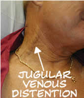
Jugular vein distention or JVD (alternately JVP — jugular vein pressure or jugular vein pulsation) is right up there among the most mentioned but least described clinical phenomena in EMS. If you tried to count how many times it occurs in your textbook, you’d run out of fingers, but many of us graduate without ever seeing so much as a picture of it, never mind developing the acumen to reliably recognize it in an emergency.

JVD is simply the visible “bulging” of the external jugular veins on either side of the neck. These are large veins that drain blood from the head and return it directly to the heart. Since they’re located near the surface, they provide a reasonably good measure of systemic venous pressure.

JVD is elevated any time venous return is greater than the heart’s ability to pump the blood back out. Remember that we’re not talking about the vessels that plug into the left heart; that involves the pulmonary arteries and veins, which are not visible in the neck. (Instead, the best indicator of pulmonary hypertension is audible fluid in the lungs.) Rather, we’re talking about the systemic vasculature, which drains into the right ventricle via the right atrium. When veins aren’t getting emptied, we look downstream to discover what portion of the pump is failing. JVD is therefore caused by right heart failure. (Of course, the most common cause of right heart failure is left heart failure, so that doesn’t mean it’s an isolated event.) If JVD isn’t the heart’s fault, then we look to fluid levels. Too much circulating volume will lead to bulging veins for obvious reasons; the flexible tubes are simply extra full.

Although it’s probably most often seen, and most diagnostic, in volume-overloaded CHF patients, the main reason JVD is harped upon in EMS is because it’s a useful sign of several acute emergencies. Mainly, these are obstructive cardiac conditions, where some sort of pressure is impeding the heart’s ability to expand, and immediate care to relieve the pressure is needed in order to prevent incurable deadness. Much like the bladder, the heart is just a supple bag of squishy muscle, and although muscle is very good at squeezing, it has no ability to actively expand. The heart therefore fills only with whatever blood passively flows into it, and if it’s being externally squeezed by pressure in the chest, it can’t fill very much.

Tension pneumothorax is perhaps the most common cause, where air leaks from the lungs into the chest cavity with no way to escape; as the pressure in the chest increases, it bears down on the heart. Associated symptoms are respiratory difficulty, decreased breath sounds on the affected side, and hypotension. Pneumothorax can be readily corrected by paramedics using needle decompression.

Cardiac tamponade is another cause, where fluid leaks from the heart into the pericardium, an inflexible sac that surrounds it (this leakage is called a pericardial effusion), eventually filling the available space and compressing the myocardium. Associated symptoms are hypotension and muffled heart sounds (these plus JVD are known as Beck’s triad). Tamponade cannot be treated in the field, but an emergency department can perform a pericardiocentesis, where a needle is inserted through the pericardium. (For the medics out there, electrical alternans on the monitor is also supportive of tamponade.)

A rather less common syndrome that can produce similar obstructive effects is severe constrictive pericarditis, inflammation of the pericardium usually caused by infection.

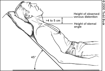
JVD is not an all-or-nothing finding — the amount of distention visible at the neck will depend on the degree of venous pressure. Gravity wants to pull blood back down, so the more venous pressure, the higher on the neck distention will climb; profound JVD reaches many inches up the neck, slight JVD will only cover a few centimeters. The pressure can actually be quantified by measuring the vertical height of the highest point of distention (measured from the heart itself, using the angle of Louis as a landmark), but this is probably more detail than is needed in the field. Suffice to say that distention reaching more than 2-4cm of vertical distance (as opposed to the distance on the neck) above the chest is usually considered pathological, and less than 1-2cm can be considered suggestive of hypovolemia.

If it changes with respiration, JVD should rise during expiration and fall with inspiration. Breathing in involves using your diaphragm to create “suction” in the chest, reducing pressure and allowing greater venous return — draining the jugulars. A paradoxical rise in JVD during inspiration (think: up when the chest goes up) is known as Kussmaul’s sign (not to be confused with Kussmaul respirations, which is a pattern of breathing), and is particularly suggestive of obstructive pathologies.

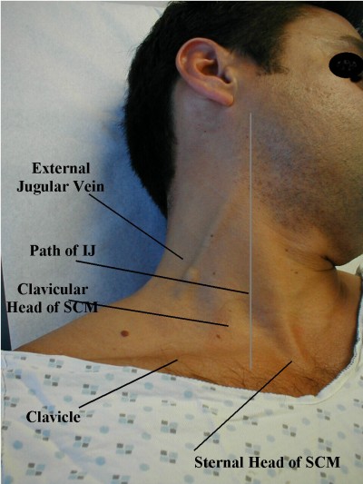
JVD can be difficult to appreciate in all but the most significant cases. It helps to turn the patient’s head away and illuminate the area with angled backlighting, which creates a “shadow” effect. Jugular pulsation should not be confused with a visibly bounding carotid pulse. To distinguish them, remember that although jugular veins may visibly pulsate, their rhythm is generally complex, with multiple pulsations for each single heartbeat (you can feel the carotid to compare the two). The jugular “pulse” will also never be palpable; the distention can be easily occluded by the fingers and will feel like nothing.

Strictly speaking, the internal jugular is usually considered more diagnostically useful than the external jugular, but it’s far harder to examine, so the latter is often used. For various reasons, many people also find the right jugular more useful than the left, although in an ambulance it’s harder to examine.

Most often, JVD is examined in an inclined or semi-Fowler’s position of 30-45 degrees. If the patient is supine, a total lack of visible JVD is actually pathological and indicative of low volume; in this position the jugular veins are usually well-filled. (Think: flat veins in a flat patient is bad.) JVD when the head is elevated is more to our interest.

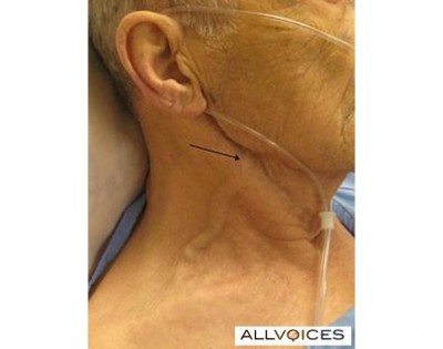
Some examples of visible JVD follow, plus some examination tips. It is recommended that you start checking this on your healthy patients now, so you’ll know what it looks like before you try to make a diagnostic call using its presence. And until you do, stop documenting “no JVD” on your assessments!

Significant JVD

Significant JVD

A different, much larger view of the same (click to enlarge)

A different, much larger view of the same (click to enlarge)

Click through for a good discussion of JVD assessment

Click through for a good discussion of JVD assessment

Some more subtle JVD

Some more subtle JVD

The basic method of measuring JVD

The basic method of measuring JVD

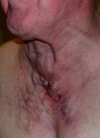
A nicely thick and squiggly external jugular

A nicely thick and squiggly external jugular

Here’s a student making her external jugular “pop” by heavily bearing down, aka the Valsalva maneuver. This markedly increases thoracic pressure, increasing venous backup; it’s an exaggeration of the effect seen during normal exhalation.

Another example of someone inducing JVD by a Valsalva

http://www.youtube.com/watch?v=FlhQGqdEfg8

Here’s a great video demonstrating the appearance of JVD, how to measure it, and testing the abdominojugular reflex (formerly known as the hepatojugular), which involves pressing down on the abdomen to raise thoracic pressure.

A brief clip of jugular venous pulsation, visible mainly toward the suprasternal notch.

http://www.youtube.com/watch?v=sOpn6_r7Wo4

Some Things to Say (part 2)


Chest pain. It’s our favorite thing to ask about and maybe our favorite thing to find. Never more does EMS get its chance to shine than when diagnosing the acute MI, and chest pain is how we start down that path. In many cases, everyone from the vomiting drunk to the elderly broken hip gets asked about their chest.

But next time you throw in, “Any chest pain?”, consider this. Not only do many heart attacks fail to present with chest pain at all, even among those that do, the specific symptoms may not amount to what your patient considers “pain.”

Pain means different things to different people. What I call pain, you might call discomfort, and my girlfriend might call a funny feeling. Tightness, palpitations, burning. Trying to list it all would leave you on scene for 20 minutes with a thesaurus, but if you don’t find the right words, then the answer you get might simply be “no.” And you’ll miss the big one.

The solution is in one magic phrase:


How does your chest feel?

I learned this gem from Captain Kent Scarna of Boston EMS, and it joins the ranks of the most useful assessment tricks out there. Because despite all the ambiguity in the chest, this one pretty much captures it all. If there’s frank pain, the patient will tell you all about it. But if there’s fluttering, itching, a feeling like they just ate a canary, this invokes that too. As a diagnostic screening, it is appropriately vague. There is a time and a place for direct questions, but when it comes to chest pain, starting off open-ended is the way to go.

How does your chest feel? Fine, it feels fine. Okay then. If you’re truly concerned you can follow up to confirm — “No pain or discomfort?” — but there’s no need to break out the Webster’s. It’s sensitive but specific; it casts a wide net, but it still unpacks fully. What else could we want?

More things to say in part 3

Managing STEMI Mimics in the Prehospital Environment

Here’s one for the medics in the audience, or anyone interested in the box with the squiggly lines.

ST elevation means acute MI. Or does it? Most medics understand that this isn’t always the case, but many don’t recognize how often it’s not, and looking deeper — sorting out true STEMI from the many non-MI pathologies that also produce ST-elevation — is not the easiest task.

The following is a PowerPoint presentation I produced for use in either continuing education coursework or merely as a standalone reference. The main intended audience is EMS providers at the paramedic level, but most of the information is at least somewhat relevant to all levels of care.

The topic is the recognition and management of “STEMI mimics,” to steal Tom Bouthillet‘s phrase: non-ACS conditions that nevertheless present with signs and symptoms resembling acute MI, particularly ECG changes such as ST segment elevation.

This is best taught by a knowledgeable instructor, but it’s designed to be usable as a self-contained reference for ambitious students; these are info-rich slides, not just graphical accompaniments to a lecture. It does assume a foundational paramedic-level education, as well as a basic understanding of ideas like sensitivity and specificity — a review of our tutorial may be in order. The illustrative ECGs are labeled with “answers” in the slide notes of the PowerPoint versions, although not on the PDF, so that’s probably the best version if this material is new to you.

Although fairly comprehensive, it’s intended as a practical guide for field assessment and treatment, rather than an in-depth examination of the etiology and course of care for every pathology discussed. For additional information, the sources for most of the contentious claims and data are listed on the slides; sources for the more everyday material are available by request. And remember to follow your service’s protocols and understand exactly where and how far you have flexibility to make some of these calls; in many cases, the decisions will be made for you.

There are 192 slides in the full presentation; the most common feedback is that this can make for a very dense and potentially drawn-out class. There is one natural “intermission” point for a break at about the halfway mark, between the introductory discussion on general ideas and before diving into specific and individual mimics. If desired, the course can be broken up further into multiple units or even multiple days.

Feel free to share, redistribute, or use for your own purposes; this is educational material made available without charge or obligation.

[Edit 10/28/12] This presentation was later enhanced into a narrated video lecture