Live from Prospect St: The Reluctant Tumble (conclusion)

Previously part 1 and part 2

Being reluctant to force Joe into an undesired ambulance ride, the crew contacted their supervisor. He arrived, evaluated the patient, agreed with their conclusions, and called Dr. Scrubs to discuss the matter. He was unable to dissuade the doctor from his decision.

The crew and supervisor approached Joe together and informed him of the circumstances; although all parties agreed that he should rightly be able to refuse transport, they felt they had been overruled by a higher authority, and if he would not come voluntarily they would be forced to compel him. Under this duress, Joe finally agreed to be transported, loudly and vocally protesting.

He was taken to his preferred hospital and care was handed off to staff with a full description of the situation. Less than 30 minutes later, another crew was sent back to the hospital to return Joe home; the attending ED physician had deemed his involuntary hold to be invalid and inappropriate, and refused to hold him against his will. No further evaluation was performed.

The encounter was documented extensively and quality improvement measures involving EMS and the base physician are expected.


Discussion

This case was not medically complicated, but it involved some difficult issues of consent and risk. Let’s look at the medicine and then at the wrinkles.

Medical Considerations

We were dispatched for a chief complaint of a fall — a very common mechanism of injury. When evaluating the fall, what should our main concerns be?

First, we should examine the mechanism itself. How far was the fall? In this case, as it often is, the fall was from a standing height, and from a standstill (i.e. not propelled while running, stumbling while breakdancing, etc.). This is often seen as the dividing line for significant versus non-significant falls; in many areas, falls from standing height or greater are considered an indication for spinal immobilization. (Other areas say greater than standing height; 3x standing height or more; or other numbers.) The elderly in particular are considered at higher risk for spinal injury, due to weakened bones and tighter ligamentous connections between vertebrae.

Typically, a blow to the head with loss of consciousness is also considered high risk for spinal injury. This is under the assumption that a blow with enough force to cause LOC may also have enough force to damage the spine. These considerations are all valid, but should only be seen as some of the many factors involved in stratifying risk; they must be considered alongside other elements like the physical assessment. In some systems, you may be forced to immobilize based on mechanism without other considerations. In others, you may be allowed to rule out immobilization based on certain findings, most of which Joe has; for instance, he denies neck or back pain or tenderness, denies peripheral parasthesias (numbness or tingling) or weakness, ambulated well, turns his head, and has no confounding factors like a distracting injury or altered mental status. In any case, the post-fall presentation was so benign that risk seemed low, and given the patient’s overall reluctance it is highly unlikely that he would have consented to a collar and board.

The use of warfarin (trade name Coumadin), on the other hand, does significantly increase the risk of intracranial hemorrhage (ICH), especially after blunt trauma to the head. Although again, Joe’s assessment was very reassuring — normal vitals, no complaints, and a baseline neurological status — it is very possible for ICH to have a delayed onset of presentation. The best example of this is the subdural hematoma, where cases of moderate severity sometimes take hours or days to develop, due to the venous rather than arterial source of bleeding. This delay is particularly common in the elderly, where (possibly due to shrinking of the gray matter, which leaves additional room for blood to collect before pressure begins compressing the brain) a classic scenario is the fall with a blow to the head, no complaints for hours afterward, and then sudden deterioration. Some sources state that 60% of geriatric fall patients who experience LOC from a blow to the head will eventually die as a result. Since in this case, we were delayed on scene for quite some time, there would be value in ongoing and repeated assessments of symptoms, neurological status, and vital signs while we waited around.

The patient’s pupils were unusual in appearance, which can be an indicator of brain herniation; however, this syndrome typically presents with one very large and round pupil. An irregularly shaped pupil as we saw here is more indicative of a structural defect, the most common of which is probably cataract surgery, which can leave the pupil off-round.

An incomplete medical history is common in scene calls involving the elderly. However, many do carry med lists, and in most cases you can reconstruct the majority of the patient’s diagnoses based on their medications. In this case, we found digoxin (or digitalis), which is almost always used to control atrial fibrillation; this is consistent with the patient’s irregular pulse, and with the warfarin, which helps prevent A-fib induced clots. Metformin (Glucophage) is an antidiabetic that helps control glucose levels. Citalopram (Celexa) is a common antidepressant of the SSRI type. Advair (fluticasone and salmeterol) is a preventative asthma/COPD inhaler combining a steroid with a long-acting beta agonist; it is used regularly to minimize flare-ups and is not a rescue inhaler. Omeprazole (Prilosec) is used for gastroesophageal reflux disease (GERD), aka heartburn. Ibuprofen is a non-steroidal anti-inflammatory (NSAID) used for pain relief and reduction of inflammation.

As VinceD noted in the comments, one essential question in any fall — and indeed in almost any traumatic event — is what caused it. Here we have a somewhat vague account which suggests a mechanical fall, i.e. tripping or loss of balance; this is not necessarily benign, as a history of repeated mechanical falls suggests deteriorating coordination or strength, but it is usually not indicative of an acute medical problem. However, many elderly patients (and some of the younger ones, too) will attribute any fall to tripping, so this claim should be taken with a grain of salt. It helps to have a witness to the event, as we do here, although witnesses are not always reliable either. In any case, what we want to know is: what happened just before the fall? Was the patient simply walking and tripped on a rug? Did he have seizure-like activity? Was he standing normally when he suddenly lost muscle tone and collapsed? Did he complain of feeling faint or dizzy? Was he exerting himself or straining on the toilet? Things happen for a reason.


Ethical and Legal Considerations

The bigger question is whether it’s okay for Joe to refuse transportation.

This is an odd question, because ordinarily we assume that people are free to go where they want, and calling 911 (or having it called for them) does not surrender this right. However, there is an attitude among those with a duty to act, such as healthcare providers and public safety officers, that individuals who are not cognitively able to understand their situation and make decisions in their best interest need to be protected from their own impaired judgment. This is equivalent to taking your friend’s keys so he won’t drive drunk, under the assumption that he wouldn’t want to drive drunk were he making sensible decisions. The legal term is implied consent, the same principle by which we transport children, drunks, and unconscious people.

How do we know if somebody is unable to make their own decisions? There is not an obvious line. For many providers, their rule of thumb is the old “A&Ox4”: if someone knows who they are, where they are, when it is, and what’s going on, then they are alert and oriented and capable of making decisions. Of course, this is only one piece of the mental puzzle. Social workers, psychiatrists, and other specialists have a full battery of tests that can help further reveal cognitive capacity. Can you perform these in the field? It’s probably more than you’re likely to do, although you might perform something simple like the MMSE. But some basic questions that highlight the patient’s judgment can help supplement your routine assessment — questions like, “Suppose you were at the mall when you started to smell smoke and heard the fire alarm. What would you do?” where any rational response is acceptable.

It’s important for the patient to be able to demonstrate that they understand what’s going on. Even someone with ordinary mental competence — unless they’re a fellow knowledgable healthcare professional — needs to be informed (to the best ability of the provider) of the possible risks and consequences of refusing care. In this case, it would involve giving them some description of the above possibilities (spinal fracture, head bleed, etc.), and ideally having the patient then relate them back to you, demonstrating good comprehension of those facts. The base physician’s view that Joe hadn’t fully demonstrated this understanding was a key part of his decision that he needed to be transported against his will.

Other important points are to ensure that the patient knows that refusal doesn’t preclude future care (“if you change your mind, you can always call back”); and that the ability of the providers to evaluate the patient on scene is at best limited. Any implication that you know what’s really happening to the patient or can definitively rule in or rule out any medical problem is unwise and legally risky. In fact, even suggesting possibilities or probabilities can be problematic if you’re wrong; on the other hand, failing to do so can leave them uninformed, so this can be a Catch 22. Your best bet is to outline some basic possibilities, carefully inform them of the limits of your training and resources, and be smart enough that you generally know what you’re talking about in the first place.

One complication in this case is the presence of someone who claims to be Joe’s health care proxy. A proxy (closely linked to the idea of a durable power of attorney) is a person whom, while of sound mind, you designate to make decisions for you if at a later time you are not of sound mind. Crucially, if you are still capable of decision-making, a proxy does not have the ability to override you; their role is to act on your behalf when you cannot. In other words, the decision of Joe’s proxy is only relevant if we do find (or in some areas, if an authority such as a judge has decided) that he’s incompetent to refuse or consent to treatment; thus, her presence does not necessarily alter the basic dilemma.

In this case, the physician’s attitude was that the problem was primarily medical: does the patient need emergency department evaluation to rule out dangerous processes? Medically, he does. However, the first question actually needs to be: Is the patient capable of evaluating risk and making decisions in his own best interest? If he is, then he is technically “allowed” to decide whatever he wants. Even a clearly dying man can refuse medical care based on religious views, personal preference, or any reason whatsoever (although barring a proxy or advanced directive, once he’s unconscious he can usually be treated under implied consent). This is different from the person who actively tries to take his own life; for philosophical reasons we view this as different from passively allowing oneself to die for lack of medical treatment. We prevent people from committing suicide but allow them to refuse medical care.

Realistically, although this fundamental right does not change, it’s fair to consider the surrounding medical circumstances to help decide how pressing and high-risk the matter is. In this case the doctor clearly felt that the risk was so high that it required going to extraordinary lengths, including overruling the patient’s own decisions and potentially even harming him, to ensure that a dangerous situation wasn’t “missed” — in short, that the ends justified the means. Dr. House is famous for this approach.

Legally, in most areas EMS providers are seen as operating under the bailiwick and legal authority of their medical director, and online medical control is an extension of this authority. In other words, within reason we are bound by the orders of medical control. The details of this relationship vary, and are not always fully explored. For an example, consider this true story from 1997 in New Jersey:

A North Bergen dual-medic crew is dispatched to a pregnant, full term female in cardiac arrest. Downtime is unknown, and they work the code for a number of minutes without response. Determining that the mother is likely unsalvageable, and concerned for the health of the fetus, they contact medical control. After a “joint decision” the base physician verbally talks them through performing an emergency C-section on scene. They deliver and successfully resuscitate the fetus, and both patients are transported. The mother is declared dead soon afterwards, but the infant lives for a number of days before dying in the hospital. In the aftermath, the paramedics are cited for violating their scope of practice, and their licenses to practice are revoked in the state of New Jersey. The physician is forced to undergo remediation training to maintain his medical control privileges.

Is the moral that acting in the patient’s best interest is not always a defense against liability? Maybe. Is the moral that medical control cannot authorize you to perform otherwise illegal acts? Maybe. Is the moral that we should protect ourselves before the patient? I don’t know about that, but it’s something to think about. In this case, the course for Joe that seems most ethical to me — allowing the patient to make his own decisions — also lets us avoid potential liability for battering and kidnapping. However, it does force us to refuse a direct order from medical control. Invoking our supervisor gives us a bigger boat either way, and would be a big help to protect us from trouble coming from our employer, one of the most likely sources. It’s also true that, while we may have believed that Joe was competent, he is at least somewhat diminished, so we’re less than completely confident. Nobody wants to put themselves on the line by taking a stand, only to be proven wrong.

Fortunately in this case we were able to avoid getting violent at all, but it was a near thing. If it did prove necessary, it should have been done with ample manpower and many hands; in some areas chemical sedation by paramedics may also be authorized. And I would certainly not recommend acting without the doctor’s signature on a legal document.

With everything viewed in retrospect, the situation would have been much more easily resolved had the doctor not been involved in the process. At the same time, however, if a simple refusal had been accepted, and CQI later went over the call — especially if Joe experienced a bad outcome — the crew would have been in a difficult place.

No matter what, such a situation is highly unusual, flush with liability, and should be thoroughly documented in all respects.

Live from Prospect St: The Reluctant Tumble

It’s 9:00PM on a Wednesday, and you’re the tech on A48, a dual-EMT, transporting BLS ambulance. You are the 911 coverage for Poketown, a midsize urban area; ALS is available for intercept as needed. You carry fingerstick glucometry, activated charcoal, glucose, aspirin, and epinephrine.

You are just starting to yawn when a tone hums from the radio, and a voice declaims:

Ambulance 8, take the response to 91 Eastbrook Rd. That’s priority 1 to 9-1 Eastbrook Road in Poketown, apartment 710, for the fall.

You acknowledge, flip your lights on, and head that way. This is an apartment block in the middle of town that you know well.

You arrive to find Poketown Fire and Police already on scene. You load your bags into the stretcher, plus a backboard, and head into the elevators, which are so small you have to fold the cot to fit inside; you wonder how you’re going to fit the patient if you end up boarding them.

You arrive at the apartment to find an elderly man sitting in his wheelchair, accompanied by neighbors and friends, including a young woman who describes herself as his healthcare proxy. He greets you cheerfully, telling you that he’s Joe, 79 years old. He was walking around the apartment with his walker when he brushed against the refrigerator and fell backwards; his proxy tried to catch him but failed, and he hit the ground. He denies falling, then denies hitting his head, saying he landed on his butt, then finally agrees that he hit his butt then his head; his proxy, however, tells you he fell straight back like a board and struck the posterior of his head on the ground. She says his eyes rolled back for a few seconds and he seemed unconscious, after which he quickly came around and moved himself to his chair. She was alarmed and called 911 immediately after; the fall was about 15 minutes ago.

He presents as fluidly conversational, friendly, and fully oriented. He is slightly hard of hearing, speaks in a loud voice, and doesn’t always understand your questions the first time around, but he’s generally “with it” and remembers the full chain of events that led him here. He jokes around with you and the firefighters and offers to marry you to one of his daughters, who has “lots of money.” You tell him you wouldn’t know what to do with it.

Physically, he seems well, with no notable trauma. There is a small lump on his occiput which may or may not be baseline (hey, heads have funny shapes), but he denies any pain or tenderness there. He also denies pain or tenderness of the neck or back, and in fact denies everything, saying he’s just fine. A “lifeline” bracelet is present on his right wrist. His vitals show:

Skin: Slightly cool in the feet, some ecchymosis throughout, otherwise dry and unremarkable.
Pulse: Weak, slightly irregularly irregular radial pulses at 78
BP: 110/70
RR: 16 and unlabored
BGL: 124

Your physical exam notes no other gross trauma. His left pupil is large and abnormal in shape; he states that he has bilateral cataracts. His right pupil is round, slightly small, and somewhat reactive. His eyes track in all directions with no major nystagmus. His lungs are clear bilaterally. He demonstrates equal CSM in all extremities, and no facial droop, arm drift, or speech slurring. A full neuro exam notes no deficits. He denies chest pain, dyspnea, nausea/vomiting, general weakness or dizziness, peripheral weakness or parasthesias (numbness/tingling), or any other complaints. During your exam, he actually gets up and ambulates back and forth across the room with his walker, moving slowly but well with no major gait disturbances.

While you talk, your partner is examining the medication list provided by his proxy. It includes:

  • Digoxin
  • Metformin
  • Citalopam
  • Advair
  • Omeprazole
  • Coumadin
  • Ibuprofen

His full medical history is otherwise not readily available. He does state that he was just released from the hospital two days ago, after a 5-day stay for diverticulitis. He is allergic to morphine.

What is your general impression of this patient’s priority?
What do you think is going on? What are you worried could be going on?
What is your next step?

Some Things to Say (part 2)


Chest pain. It’s our favorite thing to ask about and maybe our favorite thing to find. Never more does EMS get its chance to shine than when diagnosing the acute MI, and chest pain is how we start down that path. In many cases, everyone from the vomiting drunk to the elderly broken hip gets asked about their chest.

But next time you throw in, “Any chest pain?”, consider this. Not only do many heart attacks fail to present with chest pain at all, even among those that do, the specific symptoms may not amount to what your patient considers “pain.”

Pain means different things to different people. What I call pain, you might call discomfort, and my girlfriend might call a funny feeling. Tightness, palpitations, burning. Trying to list it all would leave you on scene for 20 minutes with a thesaurus, but if you don’t find the right words, then the answer you get might simply be “no.” And you’ll miss the big one.

The solution is in one magic phrase:


How does your chest feel?

I learned this gem from Captain Kent Scarna of Boston EMS, and it joins the ranks of the most useful assessment tricks out there. Because despite all the ambiguity in the chest, this one pretty much captures it all. If there’s frank pain, the patient will tell you all about it. But if there’s fluttering, itching, a feeling like they just ate a canary, this invokes that too. As a diagnostic screening, it is appropriately vague. There is a time and a place for direct questions, but when it comes to chest pain, starting off open-ended is the way to go.

How does your chest feel? Fine, it feels fine. Okay then. If you’re truly concerned you can follow up to confirm — “No pain or discomfort?” — but there’s no need to break out the Webster’s. It’s sensitive but specific; it casts a wide net, but it still unpacks fully. What else could we want?

More things to say in part 3

Pulse Oximetry: Application

The final part of a series on oximetry: start with Respiration and Hemoglobin and Pulse Oximetry: Basics

Pulse oximetry is not always available in EMS — depending on level of care, scope of practice in your area, and how your service chooses to equip you — but when it is, it’s a valuable tool in your diagnostic toolbox. Just like we discussed before, and just like any other piece of the patient assessment, using it properly requires understanding how it works and when it doesn’t.


Clinical context: When a sat is not a sat

Simply put, oximetry is the vital sign of oxygenation. It is the direct measurement of the oxygen in your bloodstream. It does not quite measure the oxygen that is actually available to your cells, but it gets close.

First, remember that actual oxygen delivery requires not just adequate hemoglobin saturation, but also enough total hemoglobin, moving around at an adequate rate. In hypovolemia, such as the shocky trauma patient, or in anemia, you might see a high SpO2 — which may be entirely accurate — but this doesn’t necessarily mean that the organs are not hypoxic. After all, you could have nothing but a single lonely hemoglobin floating around, and if it had four oxygen bound to it, you would technically have a sat of 100%. But that won’t keep anyone alive. Evaluating perfusion is a separate matter from evaluating oxygenation.

Second, remember our discussion of the oxyhemoglobin dissociation curve. The fact that you have oxygen bound to your hemoglobin doesn’t mean that it’s actually being delivered to your cells. That is, you can be hypoxic — inadequate cellular oxygenation of your organs — without being hypoxemic — inadequate oxygen present in the blood. Oximetry will only reveal hypoxemia.

Two of the strongest confounders here are cyanide and carbon monoxide (CO) poisoning. The main effect of cyanide is to impair the normal cellular aerobic cycle, preventing the utilization of oxygen; since it has no effect on your lungs or hemoglobin, the result is a normal saturation, yet profound hypoxia, since none of the bound oxygen can actually be used. Carbon monoxide, on the other hand, involves a twofer; it binds to hemoglobin in the place of oxygen, creating a monster called carboxyhemoglobin. CO has far more affinity for carboxyhemoglobin than oxygen does, so it’s hard to dislodge, and you therefore lose 1/4 of your available binding sites in the affected hemoglobin. But it doesn’t stop there. Carboxyhemoglobin also has a higher affinity for oxygen. This creates a leftward shift in the oxyhemoglobin dissociation curve — the oxygen that actually does bind finds itself “stuck,” and these well-saturated boats happily sail past increasingly hypoxic tissues without ever unloading their O2.

Consider the oximetric findings in these patients. The cyanide patient will have unimpaired blood oxygenation, so (unless he has already succumbed to respiratory failure due to the effects), a normal sat will be seen; however, hypoxia will be clinically apparent, particularly as ischemia of the heart and brain. Carbon monoxide, on the other hand, will reveal a normal or elevated (100%) sat which is partially accurate — some of that is true oxygen — and partially baloney, since CO looks the same to the oximeter as O2. But this is moot, because neither the bound CO nor the bound O2 is available to the cells. Oximeters do exist that can detect the presence of carboxyhemoglobin, known as CO-oximeters, but they are expensive and uncommon, and there is some question as to their accuracy. Your best helper here is in the patient history: both CO and cyanide are produced by fires, or any combustion in enclosed spaces (such as stoves or heaters), cyanide being released by the combustion of many plastics. You should be very wary of normal sats in any patient coming from a house fire or similar circumstances.

(Both cyanide and CO poisoning are known for causing bright red skin. In both cases oxygen is not being removed from hemoglobin, so arterial blood remains pink and well-saturated. Carboxyhemoglobin itself is also an unusually bright red. This skin, a late sign, is usually seen in dead or near-dead patients.)

Third, consider that although oximetry is an excellent measure of oxygenation, this is not the same as assessing respiratory status. It’s a little like measuring the blood pressure: although it’s a very important number, BP is an end product of numerous other compensatory mechanisms, and a normal pressure doesn’t mean that there aren’t challenges being placed on it — merely that they’re challenges you’re currently able to compensate for. Perhaps you’re satting 98%, but only by breathing 40 times a minute, and you’re fatiguing fast. Perhaps you’re satting 94%, but your airway is closing quickly and in a few minutes you won’t be breathing at all. These are clinical findings that may not be revealed in SpO2 until it’s too late.

Fourth: oximetry measures oxygenation, but not ventilation. When you breathe in, you inhale oxygen; when you breathe out, you exhale carbon dioxide. Although we use the term ventilation to describe the overall process of breathing, formally in the respiratory world it refers to the removal of carbon dioxide. Is oxygenation the more important of these two functions? Certainly; it will kill you much faster. But hypercapnia (high CO2) caused by inadequate ventilation is also a problem, and pulse oximetry does not measure it. (Capnography is the vital sign of ventilation, but that’s a topic for another day.) Now, insofar as oxygenation is primarily determined by respiratory adequacy (rate, volume, and quality of breathing), and respiration both oxygenates and ventilates, oximetry can be a good indirect measurement of ventilation; if you’re oxygenating well, you’re probably ventilating well too. This remains true if breathing is assisted via BVM, CPAP, or other device. But this is not true if supplemental oxygen is applied. Increasing the fraction of inspired oxygen (FiO2) improves oxygenation without affecting ventilation; on 100% oxygen I might be breathing 8 times a minute, oxygenating well, but ventilating inadequately.

Finally, it’s worth remembering that once you reach 100% saturation, PaO2 may no longer correlate directly with SpO2. If you reach 100% saturation at a PaO2 of 80, we could keep increasing the available oxygen until you hit a PaO2 of 500, but your sat will still read 100%. So without taking a blood gas, we don’t know whether that sat of 100% is incredibly robust, or is very close to desatting. (That’s not to say that a higher PaO2 is necessarily better; recent research continues to suggest that hyperoxygenation is harmful in many conditions. Not knowing the true PaO2 can be problematic in either direction.)


Hardware failure: When a sat is not anything

In what clinical circumstances does oximetry tend to fail? The primary one is when there isn’t sufficient arterial flow to produce a strong signal. This can be systemic, such as hypovolemia — or cardiac arrest — or it can be local, such as in PVD. (The shocked patient has both problems, being both hypovolemic and peripherally vasoconstricted.) Feel the extremity you’re applying the sensor to; if it’s warm, your chances of an accurate reading are good. The best confirmation here is to watch the waveform; a clear, accurate waveform is a very good indicator that you have a strong signal.

Tremors from shivering, Parkinsonism, or fever-induced rigors can also produce artifact on the oximeter. Some patients also just don’t like the probe on their finger. Try holding it in place, keeping the sensor tightly against the skin and the digit motionless. If there’s no luck, try another site. Any finger will work, or any toe, or an earlobe. (Some devices don’t require “sandwiching” the tissue, and can be stuck to the forehead or other proximal site, but these are uncommon in outpatient settings.)

There are a few other situations that can interfere with normal readings. In most cases, nail polish is not a problem, but dark colors do decrease the transmittance, so some shades have been reported to produce falsely low readings in the presence of already low sats or poor perfusion — as always, check your waveform for adequate signal strength. Very bright fluorescent lights have been reported to create strange numbers, and ambient infrared light — such as the heat lamps found in neonatal isolettes — can certainly create spurious readings. A few other medical oddities fall into this category as well, including intravenous dyes like methylene blue, and methemoglobinemia, which produces false sats trending towards 85%.

Is oximetry a replacement for a clinical assessment of respiration, including rate, rhythm, subjective difficulty, breath sounds, skin, and relevant history? Absolutely not. But since none of those actually provide a quantified assessment of oxygenation, they are also no replacement for oximetry. It is a valuable addition to any diagnostic suite, particularly to help in monitoring a patient over time, as well as for detecting depressed respirations before they become clinically obvious — especially in the clinically opaque patient, such as the comatose. When it’s unavailable in the field, we readily do without it. But when it’s available, it’s worth using, and anything worth using is worth understanding.

Pulse Oximetry: Basics

Just tuning in? Start with Respiration and Hemoglobin, or continue to Pulse Oximetry: Application

Once upon a time, the only way to measure SaO2 was to draw a sample of arterial blood and send it down to the lab for a rapid analysis of gaseous contents — an arterial blood gas (ABG), or something similar. This result is definitive, but it takes time, and in some patients by the time you get back your ABG, its results are already long outdated. The invention of a reliable, non-invasive, real-time (or nearly so) method of monitoring arterial oxygen saturation is one of the major advances in patient assessment from the past fifty years.

Oximetry relies on a simple principle: oxygenated blood looks different from deoxygenated blood. We all know this is true. If you cut yourself and bleed from an artery — oxygenated blood — it will appear bright red. Venous blood — deoxygenated — is much darker.

We can take advantage of this. We place a sensor over a piece of your body that is perfused with blood, yet thin enough to shine light through — a finger, a toe, maybe an earlobe. Two lights shine against one side, and two sensors detect this light from the other side. One light is of a wavelength (infared at around 800–1000nm) that is mainly absorbed by oxygenated blood; the other is of a wavelength (visible red at 600–750nm) that is mainly absorbed by deoxygenated blood. By comparing how much of each light reaches the other side, we can determine how much oxygenated vs. deoxygenated blood is present.

The big turning point in this technology came when “oximetry” turned into “pulse oximetry.” See, the trouble with this shining-light trick is that there are a lot of things between light and sensor other than arterial blood — skin, muscle, venous blood, fat, sweat, nail polish, and other things, and all of these might have differing opacity depending on the patient and the sensor location. But what we can do is monitor the amount of light absorbed during systole — while the heart is pumping blood — and monitor the amount absorbed during diastole — while the heart is relaxed — and compare them. The only difference between these values should be the difference caused by the pulsation of arterial blood (since your skin, muscle, venous blood, etc. are not changing between heartbeats), so if we subtract the two, the result should be an absorption reading from SaO2 only. Cool!

Most oximeters give you a few different pieces of information when they’re applied. The most important is the SaO2, a percentage between 0% and 100% describing how saturated the hemoglobin are with oxygen. (Typically, in most cases we refer to this number as SpO2, which is simply SaO2 as determined by pulse oximetry. This can be helpful by reminding us that oximeters aren’t perfect, and aren’t necessarily giving us a direct look at the blood contents, but for most purposes they are interchangeable terms.) But due to the pulse detection we just described, most oximeters will also display a fairly reliable heart rate for you.

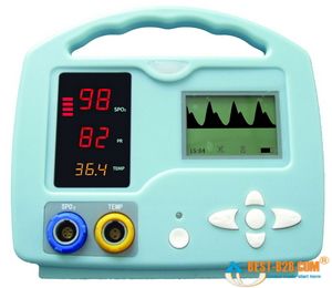
Small handheld oximeters stop there. But larger models, such as the multi-purpose patient monitors used by medics and at hospital bedsides, will also display a waveform. This is a graphical display of the pulsatile flow, with time plotted on the horizontal axis and strength of the detected pulse on the vertical. With a strong, regular pulse, this waveform should be clear and regular, usually with peaked, jagged, or saw-tooth waves. Very small irregular waves, or a waveform with a great deal of artifact, is an indicator that the oximeter is getting a weak signal, and the calculated SpO2 (as well as the calculated pulse) may not be accurate. This waveform can also be used as a kind of “ghetto Doppler,” to help look for the presence of any pulsatile flow in extremities where pulses are not readily palpable. (To be technical, this waveform is known as a photoplethysmograph, or “pleth” for short, and potentially has other applications too– but we’ll leave it alone for now.)

Most modern oximeters, properly functioning and calibrated, have an accuracy between 1% and 2% — call it 1.5% on average. However, their accuracy falls as the saturation falls, and it is generally felt that at saturations below 70% or so, the oximeter ceases to provide reliable readings. Since sats below 90% or so correspond to the “steep” portion of the oxyhemoglobin dissociation curve, where small PaO2 changes might correspond to large changes in SpO2 — in other words, an alarming change in oxygenation status — the fact that your oximeter is losing accuracy in the ranges where you most rely on it is something to keep in mind if using oximetry for continuous monitoring.

The lag time between a change in respiratory conditions (such as increasing supplemental O2 or changing the ventilatory rate) and fully registering this change on the oximeter is usually around 1 minute. And at any given time, the displayed SpO2 is a value calculated by averaging the signal over several seconds, so any near-instantaneous changes should be considered false readings.

Keep reading for our next installment, when we discuss the clinical application of oximetry, and understanding false readings.